Your Health and Fitness Partner: Androxal & FitHub

We are also excited to expand our scope by including valuable information on Androxal, a potent medication beneficial in various medical conditions. This remarkable drug, derived from the testosterone molecule, has made its mark significantly in the field of endocrinology. Patients and medical professionals can rely on our comprehensive, unbiased, and scientifically grounded content on Androxal for gaining a robust understanding of its uses, side effects, and the latest studies related to it. We understand the necessity of accurate information when it comes to medication. Our newly launched section dedicated to Androxal aims at not only educating the readers about its nuances but also at contributing beneficially to their wellbeing. Stay tuned for insightful articles unraveling the potential of Androxal in medical science.

Sitemap | Policies | Feedback    
 About the Journal
Editorial Board
Journal Subscription
Instructions for Authors
E-mail Alerts
Forthcoming Events
Advertise with Us
Contact Us
 
Article Options
FULL TEXT
PDF
Printer Friendly Version
Search Pubmed for
Search Google Scholar for
Article Statistics
Bookmark and Share
Case Report
 
Biphasic multidetector CT of primary hepatic lymphoma in a patient with HIV infection
Keywords : HIV, primary hepatic lymphoma, biphasic CT
Naveen Kalra, Sameer Vyas, Anupam Lal, Ajay Duseja, Raje Nijhawan, Niranjan Khandelwal
Departments of Radiodiagnosis and Imaging,
Hepatology and Cytology PGIMER,
Chandigarh, India, 160012.


Corresponding Author
: Dr Naveen Kalra
Email: navkal2004@yahoo.com


DOI: http://dx.doi.org/

48uep6bbphidvals|267
48uep6bbphidcol4|ID
48uep6bbph|2000F98CTab_Articles|Fulltext

Primary hepatic lymphoma (PHL) is rare in comparison to secondary liver involvement in patients with systemic lymphoma and occurs more commonly among immunocompromised patients.1 Patients usually present with nonspecific symptoms and laboratory findings. Ultrasound (US), computed tomography (CT) and magnetic resonance imaging (MRI) can detect hepatic lesions but the imaging findings are usually not specific for lymphoma.[2] A biphasic contrast-enhanced multidetector CT (MDCT) examination of the liver can exclude hypervascular lesions and helps in narrowing down the differential diagnosis. We describe the case of a patient with human immunodeficiency virus (HIV) infection and PHL and report the imaging findings on biphasic CT. US guided fine needle aspiration cytology (FNAC) of the lesion confirmed the diagnosis.
 

Case Report

A 56-year-old man with HIV infection presented with low grade, intermittent fever and dull ache in the right upper abdomen for a duration of three weeks. There was no history of jaundice, loss of appetite, loss of weight or any gastrointestinal symptoms. On examination, the liver was enlarged with irregular surface and was mildly tender on deep palpation. There was no other organomegaly and no peripheral lymph nodes were palpable. Investigations revealed haemoglobin- 11.7 m%, total leucocyte count-8400/mm3, random blood sugar-104mg%, serum amylase-50 SU, serum creatinine- 0.4mg%, bilirubin-0.6 mg%, AST- 80 IU/L, ALT-119 IU/L, Alkaline phosphatase- 186 IU/l, total serum protein- 6.9 mg/dl and CD4 count-150/µl. The peripheral blood smear was normal. Chest radiograph and contrast-enhanced CT of the chest were normal. Sonographic examination of the abdomen revealed hepatomegaly, with a large poorly marginated hypoechoic lesion in the right lobe of the liver. Biphasic contrast-enhanced multidetector CT (MDCT) of the abdomen showed a large (14 cmx10 cm), heterogeneously hypodense (5-40 HU) mass with irregular margins in segments VI and VII of the right lobe of theliver. The mass was extending laterally beyond the surface of the liver causing contour bulge. The lesion did not show any neovascularity on hepatic arterial phase (Figure 1 a&b). It was hypodense on both hepatic arterial and portal venous phase with no evidence of any contrast enhancement (Figure 2 a&b). No other lesion or lymph node was seen in the rest of abdomen. With these radiological findings, diagnosis of hepatic lymphoma or infectious aetiology were considered. US guided FNAC of the liver lesion showed low cellularity comprising predominantly of scattered immature lymphoid cells. The cells were 2-3 times the size of a mature lymphocyte with high nuclear-cytoplasmic ratio and slightly opened up chromatin. Few of the cells also showed cytoplasmic and nuclear vacuolations. In addition background showed many lymphoglandular bodies (Figure 3). A diagnosis of non- Hodgkin’s lymphoma was made. Subsequent bone marrow examination revealed no abnormality. Hence a diagnosis of PHL was made. Patient left the hospital against medical advice, before any treatment could be planned.


Discussion

PHL is a rare entity, making up to 0.4% of extranodal non- Hodgkins lymphoma, and 0.01% of all non-Hodgkins lymphoma.[1] Since 1965, only about 100 cases have been reported worldwide.[1] Secondary involvement of the liver occurs more commonly in approximately 50-60% of patients with Hodgkin’s or non-Hodgkin’s lymphoma.[2] The incidence of hepatic involvement at presentation is much lower, however occurs largely in the due course of the disease. There is increased prevalence of lymphoma among individuals with HIV infection/AIDS and the clinical presentations and CT findings are often atypical compared with lymphomas in other patients.[3] In a large biopsy proven series, the most common hepatic neoplasm in patients with AIDS was lymphoma.[4]


On imaging (USG, CT, MRI) three well recognized morphologic patterns of lymphoma have been described.These are the diffuse infiltrative variety, the nodular variety, and a mixed infiltrative and nodular variety.[5] Infiltrative involvement is most common in the secondary lymphoma, and the imaging findings may range from a normal to an  enlarged liver.[2] Hepatomegaly is however not a sensitive or a specific finding. Discrete nodular lesions are exceedingly uncommon in Hodgkin’s lymphoma, but occur in half of the patients with non-Hodgkin’s lymphoma. Nodular involvement may be detectable on imaging as single or multiple focal liver lesions.


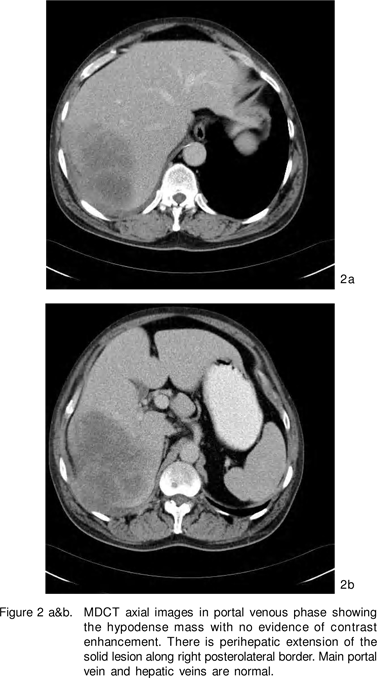
Solitary liver mass lesion is the most common presentation of PHL, followed by multiple liver lesions.[2, 6] The lesions are of low echogenicity on USG, low density on CT, and low signal intensity on T1-weighted and high signal intensity on T2- weighted MR imaging.[2, 7] Most PHLs do not show any enhancement but patchy enhancement or an enhancing ring has been described in few cases.[6, 7] Mixed infiltrative and nodular involvement has a range of imaging features of the other two varieties, such as the presence of hepatomegaly and focal liver lesions.

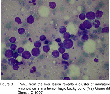
In our patient, there was no neovascularity of the lesion seen on arterial phase and no enhancement was seen on either arterial or portal venous phases of CT. The portal vein and its branches and hepatic veins were normal. As a result the hypervascular lesions like hemangioma, focal nodular hyperplasia, adenoma, hepatocellular carcinoma were excluded based on the enhancement pattern. The only differential diagnosis in a HIV positive patient based on the attenuation of the lesion and its hypovascular nature was an infectious pathology or a lymphoma. However infectious cause was less likely in the absence of thick shaggy, irregular walls of the lesion. Also there was perihepatic extension of the solid lesion without presence of any perihepatic fluid. Therefore a prospective diagnosis of a lymphoma could be made, which was confirmed on cytology.


In conclusion, biphasic MDCT helps in the diagnosis of PHL in a cases of HIV infection by showing the hypovascularity of the hepatic lesion and excluding any other organomegaly, focal lesions or lymphadenopathy.


References

1.     Avlonitis VS, Linos D. Primary hepatic lymphoma. Eur J Surg.1999;165:7259.

2.     Gazelle GS, Lee MJ, Hahn PF, Goldberg MA, Rafaat N, Mueller PR. US, CT, and MRI of primary and secondary liver lymphoma. J Comput Assist Tomogr. 1994;18:4125.

3.     Nyberg DA, Jeffrey RB Jr, Federle MP, Bottles K, Abrams DI. AIDSrelated lymphomas: evaluation by abdominal CT. Radiology. 1986;159:5963.

4.     Poles MA, Dieterich DT, Schwarz ED, , Lew EA, Lew R, et al. Liver biopsy f indings in 501 pat ients infected with human immunodeficiency virus (HIV). J Acquir Immune Defic Syndr Hum Retrovirol. 1996;11:1707.

5.     Maher MM, McDermott SR, Fenlon HM, Conroy D, O’Keane JC, Carney DN, et al. Imaging of primary non-Hodgkin’s lymphoma of the liver. Clin Radiol. 2001;56:295301.

6.     Sanders LM, Botet JF, Straus DJ, Ryan J, Filippa DA, Newhouse JH. CT of primary lymphoma of the liver. AJR Am J Roentgenol. 1989; 152:9736.

7.     Memeo L, Pecorello I, Ciardi A, Aiello E, De Quarto A, Di Tondo U. Primary non-Hodgkin’s lymphoma of the liver. Acta Oncol. 1999; 38:6558.