Lipomas of the small bowel are uncommon benign tumours; most are asymptomatic. Over 200 cases of duodenal lipomas have been reported, but presentation with severe upper GI haemorrhage is unusual and has rarely been reported.[1,2,3,4,5,6]
Case Report
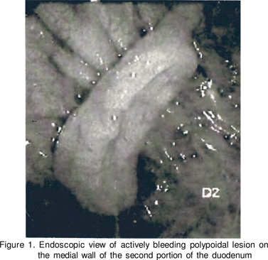
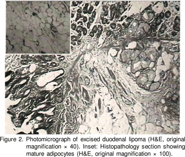
A 70-year-old woman was admitted with a history of passage of tarry stools for two day. She had no history of abdominal pain, drug/alcohol intake and had no past history of GI bleed. Physical examination demonstrated pallor, tachypnea (28/ minute), a systolic pressure of 90 mm Hg, and pulse of 120 beats per minute. The abdomen and other systemic examinations were unremarkable. Laboratory investigations revealed a haemoglobin of 7.4 g/dl, white cell count of 8800/ mm3 with normal differential count and platelet count of 230,000/mm3. Liver, renal, electrolyte, and coagulation panels were normal. Emergency upper gastrointestinal endoscopy after initial haemodynamic stabilisation demonstrated a large pedunculated lesion in the second portion of the duodenum with active oozing of blood (Figure 1). The lesion was removed by endoscopic electrosurgical snare polypectomy, and the specimen recovered. The polypoid mass measured 5.5 cm × 1.4 cm × 1 cm with ulceration at the tip. The final histopathological diagnosis was that of a lipoma (Figure 2). The patient was followed up for one year and had no recurrent bleeding.
Discussion
Lipomas have been found throughout the GI tract but occur most commonly in the colon, ileum, and jejunum.[1,7,8] Lipomas of the duodenum are relatively rare and are generally located in the second portion of the duodenum.[7] The incidence of lipomas among all benign small-bowel tumours is reported to be 21.4%.[9] In a review of 1200 consecutive duodenoscopies, lipomas were found in only 2 patients.[10] Most small intestinal lipomas are often found incidentally, and symptoms occur in less than a third of affected patients. Based on a review of published reports, it appears that most duodenal lipomas greater than 4 cm in diameter produce symptoms.[1,10] The most common manifestation is abdominal pain, followed by palpable mass, nausea, vomiting, and diarrhoea caused by intussusception. Others symptoms are related to slow gastric emptying and chronic hypochromic microcytic anaemia; the latter is usually caused by complications including intussusception, surface ulceration, and obstruction.[1] Acute upper GI haemorrhage is a rare complication. Over 200 cases of duodenal lipomas have been reported, but in fewer than 15 the presentation was severe upper GI haemorrhage.[1,2,7] Duodenal lipomas are most often submucosal, but can also be subserosal.[11] The shape is variable and they can be either sessile or pedunculated. The overlying mucosa is usually normal, but there may be areas of ulceration. The mechanisms of ulceration are probably mucosal pressure atrophy or peristalsis leading to elongation and stretching with necrosis of the overlying epithelial layers.[1,12] On barium contrast radiography duodenal lipomas can appear as pedunculated masses or as rounded filling defects within the lumen of the duodenum.[1,7,8] Computed tomography allows a definitive preoperative diagnosis because attenuation values in the range of minus 50 to 100 Hounsfield units confirm that a lesion has significant lipomatous component.[1,12,13] The typical endoscopic appearance is that of a smooth, hemispherical polypoid lesion with a wide base. However the endoscopic features of duodenal lipomas are nonspecific and deep biopsies are needed for definitive diagnosis. Endoscopic ultrasound (EUS) is considered highly useful for the diagnosis of submucosal tumours of the GI tract. EUS features of lipomas, specifically a markedly homogenous hyperechoic mass within the submucosal layer, are highly characteristic and are useful for differentiating a lipoma from a gastrointestinal stromal tumour.[14,15] For symptomatic lipomas, surgical resection has traditionally been required. However if pedunculated, as in our patient, the lipoma can be easily and safely removed by endoscopic snare polypectomy.[4,6,10] However, if the lesion is clearly sessile or the base of the tumour is large, snaring is difficult and problematic because of the risks of perforation, the need for increased coagulation current (because of the high water density of the fat), and serosal invagination of a pseudopedicle.[16].


References
1. Michel LA, Ballet T, Collard JM, Bradpiece HA, Haot J. Severe bleeding f rom submucosal lipoma of the duodenum. J Clin Gastroenterol. 1988;10:541–5.
2. Agha FP, Dent TL, Fiddian-Green RG, Braunstein AH, Nostrant TT. Bleeding lipoma of the upper gastrointestinal tract. A diagnostic challenge. Am Surg. 1985;51:279–85
3. Hamperl WD, Wagner T. Bleeding duodenal lipoma — a rare finding. Chirurg. 1990;61:331–2.
4. Tung CF, Chow WK, Peng YC, Chen GH, Yang DY, Kwan PC. Bleeding duodenal lipoma successfully treated with endoscopic polypectomy. Gastrointest Endosc. 2001;54:116–7.
5. Dargan P, Sodhi P, Jain BK. Bleeding gastric lipoma: case report and review of the literature. Trop Gastroenterol. 2003;24:213–4.
6. Sou S, Nomura H, Takaki Y, Nagahama T, Matsubara F, Matsui T, et al. Hemorrhagic duodenal lipoma managed by endoscopic resection. J Gastroenterol Hepatol. 2006;21:479–81.
7. Krachman MS, Dave PB, Gumaste VV. Bleeding duodenal lipoma [letter]. J Clin Gastroenterol. 1992;15:180–1.
8. Munson FA. Duodenal lipoma. A report of two cases with a review of the literature. Am J Gastroenterol. 1972;58:613–8.
9. Olmsted WW, Ros PR, Hjermstad BM, McCarthy MJ, Dachman AH. Tumors of the small intestine with litt le or no malignant predisposition: a review of the literature and report of 56 cases. Gastrointest Radiol. 1987;12:231–9.
10. Wald A, Milligan FD. The role of fiberoptic endoscopy in the diagnosis and management of duodenal neoplasms. Am J Dig Dis. 1975;20:499–505.
11. Reddy RR, Schuman BM, Priest RJ. Duodenal polyps: diagnosis and management. J Clin Gastroenterol. 1981;3:139–47.
12. Whetstone MR, Zuckerman MJ, Saltzstein EC, Boman D. CT diagnosis of duodenal lipoma. Am J Gast roenterol. 1985;80:251–2.
13. Waligore MP, Stephens DH, Soule EH, McLeod RA. Lipomatous tumors of the abdominal cavity: CT appearance and pathologic correlation. Am J Roentgenol. 1981;137:539–45.
14. Nakamura S, Iida M, Suekane H, Matsui T, Yao T, Fujishima M. Endoscopic removal of gastric lipoma: diagnostic value of endoscopic ult rasonography. Am J Gast roenterol. 1991;86:619–21.
15. Hizawa K, Kawasaki M, Kouzuki T, Aoyagi K, Fujishima M. Unroofing technique for the endoscopic resection of a large duodenal lipoma. Gastrointest Endosc. 1999;49:391–2.
16. Khawaja FI. Pedunculated lipoma of the colon. Risk of endoscopic removal. South Med J. 1987;80:1176–9.