|
|
|
|
 |
 |
| |
 |
|
|
Case Report |
|
|
|
|
|
Keywords :
Fasciolopsis buski, intestinal perforation |
|
|
Hemanga K Bhattacharjee, Devendra Yadav, Deepak Bagga
Department of Paediatric Surgery,
Safdarjung Hospital,
New Delhi,110029, India
Corresponding Author:
Deepak Bagga
Email:deepak_bagga2000@yahoo.co.uk
DOI:
http://dx.doi.org/
48uep6bbphidcol4|ID 48uep6bbphidvals|294 48uep6bbph|2000F98CTab_Articles|Fulltext Fasciolopsis buski is the largest fluke parasitising humans.[1] Fasciolopsiasis is seen in various parts of South-east Asia including China, Taiwan, Bangladesh, India, Vietnam and Thailand.[2,3,4,5] In India, it has been reported from Assam, West Bengal, Bihar, Uttar Pradesh and Maharashtra.[6,7,8]
Adult worms live in the small intestine of humans and pigs. Eggs are liberated in the faeces. They encyst as metacercariae in fresh water plants after passing through the intermediate host snail. Human infection occurs through peeling off of the skin or hull of raw infected plants between one’s teeth or drinking of untreated water containing the metacercariae. The worm normally inhabits the duodenal and jejunal mucosa; in moderate and heavy infection it can be found in much of the intestinal tract including the stomach and colon.[9] The pathological changes may be traumatic, obstructive or toxic. Heavy infection causes extensive intestinal and duodenal erosion, ulceration, haemorrhage, abscess and catarrhal inflammation. Absorption of toxic and allergic worm metabolites causes facial oedema and generalised ascites.[10]
Clinical features are related to the parasitic load. Most infections are mild and asymptomatic. Heavy infection causes abdominal pain, diarrhoea, flatulence, poor appetite, vomiting, intestinal obstruction, eosionophilia and leucocytosis.[4,5,8,11,12] Ascites, anasarca and death are reported in severe cases.[8,10,12]
Fasciolopsiasis is a persistent public health concern. An awareness and recognition of the entity is required for prompt patient management.
Case report
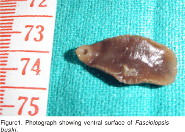
A 10-year-old boy from Barabanki district of Uttar Pradesh, India, presented with history of central abdominal pain and abdominal distension for 3 days and fever for 2 days. He was febrile, and dehydrated with tachycardia and signs of peritonitis. An x-ray of the abdomen showed multiple air fluid levels. Blood biochemistry revealed total leucocyte count of 8000/mm3, (neutophils 64, lymphocytes 32, monocytes 2, eosonophils2). After resuscitation he was taken up for emergency laparotomy. There was a perforation approximately 1 cm in diameter on the antemesenteric border of the mid ileum. The peritoneal cavity was filled with dirty coloured intestinal fluid, pus and large numbers of the leaf-shaped fleshy worms (10- 15); a few of them were alive and floating. Another 10-15 wormswere removed on milking the intestine (Figure 1).

A loop ileostomy was performed after thorough peritoneal lavage. He developed wound infection and dehiscence during the post operative period which was managed conservatively. His blood culture showed no growth and the Widal test was negative. The worm was confirmed as Fasciolopsis buski on histopathological examination. Stool sample taken from the rectum showed eggs of Fasciolopsis buski (Figure 2). On the 7th postoperative day oral praziquantel (25 mg/kg) was prescribed in three divided doses daily for one day.
Discussion
The worm load of this patient was astonishing. The child was harbouring as many as 25 worms. Thus it can be inferred that he had consumed a good many metacercariae forms over a prolonged period as there is no multiplication inside the definitive host but only development. The implication of this is the obvious paucity of preventive measures and health education despite the endemicity of the condition. Heavy infestation may cause intestinal obstruction, deep ulceration, haemorrhage and abscess on the intestinal wall. No report of fasciolopsiasis of this magnitude presenting as intestinal perforation has been made in the past. In this case there is a distinct possibility that heavy worm infestation and subsequent traumatic ulceration of the intestinal wall resulted in the perforation. Hence, paediatric patients with intestinal perforation from the endemic areas need to be looked for
fasciolopsiasis.
Reference
1. Kuntz RE, Lo CT. Preliminary Studies on Fasciolopsis buski (Lankester, 1857) (Giant Asian Intestinal Fluke) in the United States. Trans Amer Microsc Soc. 1967;86:163–6.
2. Lo CT, Lee KM. Intestinal parasites among the Southeast Asian laborers in Taiwan during 1993-1994. Zhonghua Yi Xue Za Zhi (Taipei). 1996;57:401–4.
3. Wiwanitkit V, Suwansaksri J, Chaiyakhun Y. High prevalence of Fasciolopsis buski in an endemic area of liver fluke infection in Thailand. MedGenMed. 2002;4:6.
4. Gilman RH, Mondal G, Maksud M, Alam K, Rutherford E, Gilman JB, et al. Endemic focus of Fasciolopsis buski infection in Bangladesh. Am J Trop Med Hyg. 1982;31:796–802.
5. Le TH, Nguyen VD, Phan BU, Blair D, McManus DP. Case report: unusual presentation of Fasciolopsis buski in a Vietnamese child. Trans R Soc Trop Med Hyg. 2004;98:193–4.
6. Bhatti HS, Malla N, Mahajan RC, Sehgal R. Fasciolopslasis—a reemerging infection in Azamgarh (Uttar Pradesh). Indian J Pathol Microbiol. 2000;43:73–6
7. Manjarumkar PV, Shah PM. Epidemiological study of Fasciolopsis Buski in Palghar Taluk. Indian J Public Health. 1972;16:3–6.
8. Kumari N, Kumar M, Rai A, Acharya A. Intestinal trematode infection in North Bihar. JNMA J Nepal Med Assoc. 2006;45:204–6.
9. Graczyk TK, Gilman RH, Fried B. Fasciolopsiasis: is it a controllable food-borne disease? Parasitol Res. 2001;87:80–3
10. Jaroonvesama N, Charoenlarp K, Areekul S. Intestinal absorption studies in Fasciolopsis buski infection. Southeast Asian J Trop Med Public Health. 1986;17:582–6.
11. Gupta A, Xess A, Sharma HP, Dayal VM, Prasad KM, Shahi SK. Fasciolopsis buski (giant intestinal fluke)—a case report. Indian J Pathol Microbiol. 1999;42:359–60.
12. Mas-Coma S, Bargues MD, Valero MA. Fascioliasis and other plantborne trematode zoonoses. Int J Parasitol. 2005;35:1255–78.
|
|
|
 |
|
|