|
|
|
|
 |
 |
| |
 |
|
|
Case Report |
|
|
|
|
|
Keywords :
hemangioma, liver resection, cholangiocarcinoma, liver tumour |
|
|
Disha Sood,1 Vinay Kumaran,1 TBS Buxi, 2 Samiran Nundy 1, Arvinder Singh Soin
Department of Surgical Gastroenterology & Liver Transplantation1,
and Radiology2 Sir Ganga Ram Hospital
New Delhi, India
Corresponding Author:
Dr Vinay Kumaran
Email: kumaranvinay@yahoo.com
DOI:
http://dx.doi.org/
48uep6bbphidcol4|ID 48uep6bbphidvals|297 48uep6bbph|2000F98CTab_Articles|Fulltext In spite of advances in radiological imaging, preoperative diagnosis of liver tumours is problematic. Benign lesions may mimic liver malignancies and patients may unnecessarily undergo extended resection for what may have been essentially benign. Here we present the case of a patient who underwent extended liver resection for suspected intra-hepatic cholangiocarcinoma based on preoperative radiological imaging which was later diagnosed as a hemangioma on histopathology.
Case Report
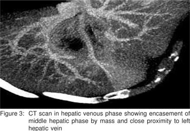
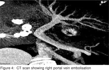
A 55-year old man presented with dyspeptic symptoms of 3 weeks’ and pain in right hypochondrium of one week’s duration. Physical examination revealed no palpable liver mass and there was no jaundice or ascites. The liver function tests including aspartate aminotransferase (40 IU/L), alanine aminotransferase (35 IU/L) and total bilirubin (1.1 mg/dL) levels were normal. Hepatitis B surface antigen and hepatitis C antibody were both negative. Alpha-fetoprotein, CEA and CA 19-9 were normal. CT angiography showed a central hypodense lesion 6.7 cm x 6.5 cm x 4.6 cm involving segments 1, 4, 5 and 8 of the liver encasing the middle hepatic vein and there were multiple hemangiomatous lesion in both lobes of the liver (Figures 1, 2, 3 and 4). Positron emission tomography scan described an ill-defined peripherally enhancing lesion measuring approximately 4 cm x 6.6 cm in segments 4 and 8, abutting the left branch of the portal vein with the surrounding fat planes maintained. Another lesion 2 cm x 1 cm was seen in segment 2, suggesting a hemangioma. A presumptive diagnosis of cholangiocarcinoma was made and we planned a right trisegmentectomy, which would have involved resecting the right lobe along with segment 4.
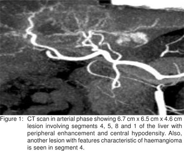
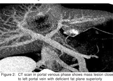
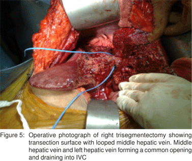
However as the calculated volume of the remaining liver i.e. the left lateral segment was only 17% (against the minimum post resection volume requirement of 25% in patients with normal livers[1]) we embolised the right portal vein. After five weeks, a repeat CT volumetric calculation showed that the left lateral segment had hypertrophied to 27% of the total liver volume and the patient was taken up for surgery. The intraoperative findings showed neither peritoneal metastases, nor ascites or satellite lesions. There was a mass however, involving segments 4, 5, 8 and 1 of the liver, and the middle hepatic vein (Figure 5). The left hepatic artery was not involved. Intraoperative ultrasound showed that the left portal and left hepatic veins were also free from the mass. The right anterior and posterior sectoral ducts were seen separately entering the bile duct. Under low CVP anaesthesia using an ultrasonic aspirator, liver transection to the left of the falciform ligament was carried out, after ligating the right hepatic artery and the right portal vein. The right hepatic vein and middle hepatic veins were divided, the latter at the point where the middle and left hepatic veins drained into the inferior vena cava. The right trisegmentectomy was completed with a caudate lobectomy. The patient had an uneventful postoperative course. The resected specimen showed a hard greyish white tumour 5 cm x 4 cm in diameter with ill-defined margins and microscopic examination revealed a tumour composed of numerous proliferating capillary-sized blood vessels surrounded by loose oedematous stroma. There was no evidence of malignancy. A pathological diagnosis of hemangioma was made. Due to preoperative portal vein embolisation the sclerosed variant could not be clearly identified.
Discussion
The commonest benign tumour of the liver is a hemangioma, which has a reported incidence of 1 to 20%.[2] The diagnosis of hemangioma on CT scan is established based on its characteristic features which include early enhancement with centripetal filling that progresses to uniform filling in the portal venous phase and the persistence of contrast in the delayed phase.[3] However this appearance may be misdiagnosed as a tumour if degenerative changes occur leading to sclerosis of the hemangioma.
Shepherd and Lee[4] were the first to describe five cases of this ‘rare’ tumour with a central necrotic component and a peripheral hyalinised fibrotic capsule. There have only been eleven cases reported in the literature[5] and sclerosed haemangioma is seen as a low-density lesion on plain CT with contrast CT showing a ring-like enhancement, which is typical of adenocarcinomas.[6,7]
The differential diagnosis includes fibrolamellar hepatocellular carcinoma, colorectal metastasis and cholangiocarcinoma, which are hypovascular adenocarcinomas.[2,7] It is difficult to differentiate the appearance on imaging from colorectal metastases. CEA measurement may be useful as a CEA level of >5 ng/ml occurs in 98% of such patients.[8] MRI shows low signal intensity on T1 and high signal intensity on T2 images, which is not characteristic of a hemangioma.[5] FDG- PET has a negative predictive value of less than 50% and sensitivity of 80% for hepatocellular carcinoma.[9]
On histopathological examination these hemangiomas show appearances ranging from capillary-sized vascular channels to cavernous channels lined with a single layer of flat endothelial cells.[10] Factor VIII related antigen is the immunohistochemical marker that can differentiate them from primary hepatocellular carcinomas and other liver metastases. In this case it was not possible to confirm the degenerative changes in the haemangioma because of the preoperative portal vein embolisation that had been carried out.
Fine needle aspiration cytology has an accuracy of 90- 93% in detecting hepatic malignancy,[11] which is comparable
to radiological investigation.[12] In patients with colorectal metastases, major surgery for suspected malignant disease, that is actually benign, has been reported in only 1.9%.[13] However, in primary liver tumours, the risk of needle tract seeding is 0.4% to 5.1%[14] and hence preoperative biopsy is not indicated if there is suspicion of malignancy and resection is proposed. Also, liver resection from experienced centres have reported mortality rates of less than 5% and less than 1% in patients with no cirrhosis.[15] Therefore when malignancy cannot be ruled out these patients should be offered surgery. Fine needle aspiration may not only provide false reassurance with a negative result but also the risk of needle tract metastasis outweighs its benefit.
Sclerosed hemangioma is a rare diagnosis, which should be considered in the differential diagnosis of liver tumours before contemplating major surgery.
References
1. Azoulay D, Castaing D, Krissat J, Smail A, Hargreaves GM, Lemoine A, et al. Percutaneous portal vein embolisation increases the feasibility and safety of major liver resection for hepatocellular carcinoma in injured liver. Ann Surg. 2000;232:665–72.
2. Goodman ZD. Benign tumour of the liver. In: Okuda K, Ishak KG, editors. Neoplasms of the liver. Berlin Heidelberg New York: Springer. 1987;105–25.
3. Nelson RC, Chezmar JL. Diagnostic approach to hemangiomas. Radiology. 1990;176:11–3.
4. Shepherd NA, Lee G. Solitary necrotic nodules of the liver simulating hepatic metastasis. J Clin Pathol. 1983;36:1181– 3.
5. Mori H, Ikegami T, Imura S, Shimada M, Morine Y, Kanemura H, et al. Sclerosed hemangioma of the liver: Report of a case and review of the literature. Hepatol Res. 2008;38:529–33.
6. Haratake J, Horie A, Nagafuchi Y. Hyalinised haemangioma of the liver. Am J Gastroenterol. 1992;87:234–6.
7. Doyle DJ, Khalili K, Guindi M, Atri M. Imaging features of sclerosed hemangioma. AJR Am J Roentgenol. 2007;189:67–72.
8. Tartter PI, Slater G, Gelernt I, Aufses AH Jr. Screening for liver metastasis from colorectal cancer with carcinoembryonic antigen and alkaline phosphatase. Ann Surg. 1981;193:357–60.
9. Ho CL, Chen S, Yeung DW, Cheng TK. Dual tracer PET/CT imaging in evaluation of metastatic hepatocellular carcinoma. J Nucl Med. 2007;48:902–9.
10. James M, Crawford MD. Pathological assessment of liver cell dyplasia and benign liver tumors: differentiation from malignant tumors. Semin Diagn Pathol. 1990;17:115–28
11. Durand F, Regimbeau JM, Belghiti J, Sauvanet A, Vilgrain V, Terris B, et al. Assessments of the benefits and risks of percutaneous biopsy before surgical resection of hepatocellular carcinoma. J Hepatol. 2001;35:254–8.
12. Kinkel K, Lu Y, Both M, Warren RS, Thoeni RF. Detection of hepatic metastasis from cancers of the gastrointestinal tract by using noninvasive imaging methods (US, CT, MR imaging, PET): a metaanalysis. Radiology. 2002;224:748– 56
13. Clayton RAE, Clarke DL, Currie EJ, Madhavan KK, Parks RW, Garden OJ. Incidence of benign pathology in patients undergoing hepatic resection for suspected malignancy. Surgeon. 2003;1:32–8.
14. Takamori R,Wong LL, Dang C,Wong L. Needle-tract implantation from hepatocellular cancer: is needle biopsy of the liver always necessary? Liver Transpl. 2000;6:67–72.
15. Belghiti J, Hiramatsu K, Benoist S, Massault P, Sauvanet A, Farges O. Seven hundred forty-seven hepatectomies in the 1990s: an update to evaluate the actual risk of liver resection. J Am Coll Surg. 2000;191:38–46.
|
|
|
 |
|
|