|
|
|
|
 |
 |
| |
 |
|
|
Case Report |
|
|
|
|
|
Keywords :
|
|
|
Amagunloye, Aoadeyinka, Ja Otegbayo
Department of Radiology,
University College Hospital,
Ibadan, Oyo State, Nigeria
Corresponding Author:
Dr. Atinuke M Agunloye
Email: tinuagunloye@comui.edu.ng
DOI:
http://dx.doi.org/
48uep6bbphidvals|214 48uep6bbphidcol2|ID 48uep6bbph|2000F98CTab_Articles|Fulltext A 72 year-old-male patient, presented to the gastrointestinal unit of the medical out-patients’ department of the University College Hospital (UCH), Ibadan, Nigeria. His complaints were unexplained weight loss of two months’ duration and abdominal pain of 1 week’s duration, felt in the epigastric and periumbilical regions. The pain was worse with meals but the patient claimed that there was no loss of appetite. There was occasional nausea but no vomiting. The patient gave no history of melena, and he did not have altered bowel habits. He was not a known hypertensive or diabetic but had a history of benign prostatic hyperplasia. Drug history revealed an occasional use of the NSAID diclofenac sodium, but he had ceased using it three months prior to presentation.
The only positive finding on physical examination was tenderness in the epigastric region with no palpable abdominal mass. A barium study of the upper gastrointestinal tract suggested chronic peptic ulcer disease. Omeprazole was prescribed and the patient reported some relief of symptoms. However, a routine abdominal ultrasound done revealed multiple rounded hypoechoic masses of varying sizes in the right lobe of the liver which were avascular on colour flow Doppler examination.
The prostate specific antigen (PSA) was moderately increased (10.1 µg/L). The hemogram was normal and he was negative for the hepatitis C and B surface antigens as well as the human immunodeficiency virus (HIV). The carcinoembryonic antigen (CEA) was significantly raised with a value of 317.8 µg/L.
An abdominal computerised tomography (CT) scan was requested based on the ultrasound findings, persistent epigastric pain and abnormal CEA value, to rule out an upper abdominal malignancy. Oral contrast was given twenty minutes before and another bolus just before the scan, as part of the routine preparation for upper abdominal CT scan at our centre. However, the CT scan machine developed a fault just before the examination and the study was not performed until about 7 hours after contrast ingestion. The entire colon was well coated with contrast at this time. Another bolus of oral contrast was given immediately before the scan to ensure distension of the stomach and small bowel.
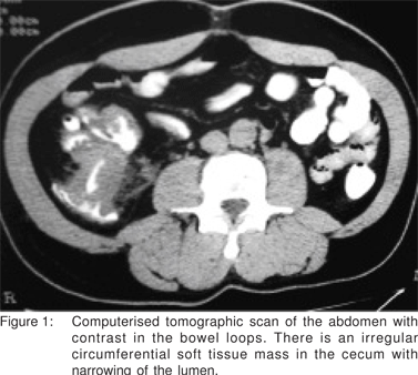
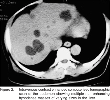
The CT scan (Figure 1) showed a cecal pole mass with irregular luminal narrowing and mucosal destruction. There were also multiple non-enhancing masses in both lobes of the liver (Figure 2). The pancreas and stomach were normal. Atherosclerotic plaques were noted in the aorta and iliac vessels. There were also lumbar spondylotic changes with facet degeneration.


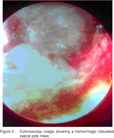
Upper and lower gastrointestinal videoendoscopy was then requested for clarification. This revealed a normal oesophagus and evidence of antral gastritis. The colon contained multiple diverticuli with a lobulated caecal pole mass (Figure 3). Biopsy specimens taken from the stomach and caecal mass were reported as chronic gastritis and adenocarcinoma, respectively.

Discussion
Cecal pole tumour is a known cause of unexplained weight loss with no other localising signs; this was also seen in the present case. The actual diagnosis may also be delayed in patients with other symptomatic coexisting diseases. Patients with distal colonic tumour may complain of diarrhoea alternating with constipation but the cecal pole, being capacious rarely presents with obstructive symptoms when it harbors a tumour. However, in elderly patients with any symptom referable to the abdomen, it is necessary to screen for occult malignancies. Abdominal ultrasound may be helpful in such cases as an initial screening tool as demonstrated in this case. Barium studies of either the upper or lower gastrointestinal tract may be requested depending on the specific suggestive symptoms present, but as seen in this case these symptoms may be deceptive. The focus of investigations in this patient was on the upper GIT as this patient was initially thought to have peptic ulcer disease.
In abdominal CT examinations, the bowel opacification technique and image acquisition are tailored to the suspected site of pathology. In our centre, oral contrast is given 20 minutes before the CT scan to opacify the small bowel while another bolus is given immediately before the CT scan to distend the stomach and duodenum. This technique is satisfactory for evaluating the upper abdominal organs and small bowel. However, this timing of contrast ingestion will normally not result in satisfactory distension of the colon. For adequate colon distension, 400-600 mL of oral contrast is given on the evening before the CT scan or 2-3 hours prior to CT scan. Water and air can also be introduced per rectum to achieve colonic distension.[1,2,3,4].
The unintended long time lapse between oral ingestion of the contrast and CT scan in our case, with resultant colonic distension was responsible for the identification of the cecal tumour which was not suspected clinically. CT also better delineated the hepatic metastatic deposits as was expected. The fact that CT allows other associated findings, both intraand extra colonic, to be obtained thus improving the diagnostic and therapeutic management of the patient is an advantage over barium enema.
Numerous studies have been done to evaluate the accuracy of the CT technique of using prolonged oral contrast medium two days prior to the CT scan, called ‘minimal preparation abdominal CT’, rather than rectal instillation which might be uncomfortable for the patient. These studies have demonstrated satisfactory results for diagnosing colonic cancers in the caecum.[5, 6] This technique is reported to be especially useful in frail, elderly and immobile patients who frequently have difficulty in tolerating conventional colonic investigations like barium enema and colonoscopy. The study by Ng et al5 demonstrated sensitivity, specificity, negative predictive values (NPV) and positive predictive values (PPV) of 90%, 98%, 99% and 56%, respectively for the minimal preparation technique in the identification of caecal carcinomas. The minimal preparation technique was somewhat inevitably achieved in this patient because of a fault in the only available CT machine which was to be used for the study. We realise that we might have missed the cecal tumor in this patient with our protocol, and the diagnosis may thus have been further delayed.
In the study by Domjan[6] and his colleagues, of the 118 elderly patients with suspected colonic pathologies, a comparison of CT and BE was made and both techniques congruently gave negative reports in 68.8% of patients. Hence, radiologists and referring clinicians should be aware that colonic lesions may still be occasionally missed on CT but the study has the additional advantage that other significant pathologies unrelated to the colon might be detected in other abdominal organs as was seen in this patient’s liver. CT is also useful in tumour staging, as tumour invasion beyond the bowel wall and nodal metastases can be demonstrated.
Recently, because of its fast and thin-section scanning abilities, virtual colonoscopy with multidetector-row CT (MDCT) is said to be an efficient screening method for colon cancer, and MDCT enterography is becoming the standard imaging technique for many small bowel disorders.[1] MR colonography (MRC) is also reported to be an accurate diagnostic tool for the detection of colorectal masses and inflammatory diseases.[7]
Colonoscopy, if available and where a complete examination is achieved, is the current gold standard procedure as it is more sensitive than other tests for the detection of colonic cancers and polyps.[8] Colonoscopy was confirmatory in this case and enabled tissue samples to be taken for histological diagnosis.
In conclusion, the authors recommend that in elderly patients with unexplained weight loss and anaemia with associated abdominal symptoms, a single CT scan of the entire abdomen following ingestion of oral contrast medium, 6-24 hours before the scan, may be considered as one of the imaging options. This will help diagnose upper or lower GIT mass lesions and/or other abdominal pathologies and save expenses especially in resource-poor settings.
References
1. Erturk SM, Mortele KJ, Oliva MR, Barish MA. State of the art computed tomographic and magnetic resonance imaging of the gastrointestinal system. Gastrointest Endosc Clin N Am. 2005;15:581–614.
2. Hyun Kwon Ha, Kim JK.The Gastrointestinal Tract. In: CT and MR Imaging of the whole body, edited by Haaga J R, Lanzieri CF and Gilkeson RC. 4th Ed. Mosby Inc, USA.2003 pp 1154–5.
3. Parlorio de Andrés E, Soria Aledo V, Pellicer Franco E, Flores Pastor B, Morales Cuenca G. Computed tomographic colonography: applications, advantages and disadvantages. Gastroenterol Hepatol. 2005;28:365–8.
4. Angelelli G, Macarini L. CT of the bowel: use of water to enhance depiction. Radiology. 1988;169:848–9.
5. Ng CS, Doyle TC, Pinto EM, Courtney HM, Miller R, Bull RK, et al. Caecal carcinomas in the elderly: useful signs in minimal preparation CT. Clin Radiol. 2002;57:359–64.
6. Domjan J, Blaquiere R, Odurny A. Is minimal preparation computed tomography comparable with barium enema in elderly patients with colonic symptoms? Clin Radiol. 1998;53:894–8.
7. Lauenstein TC, Kuehle CA, Ajaj W. MR imaging of the large bowel. Magn Reson Imaging Clin N Am. 2005;13:349–58.
8. Rockey DC, Paulson E, Niedzwiecki D, Davis W, Bosworth HB, Sanders L, et al. Analysis of air contrast barium enema, computed tomographic colonography, and colonoscopy: prospective comparison. Lancet. 2005;365:305–11.
|
|
|
 |
|
|