|
|
|
|
 |
 |
| |
 |
|
|
Case Report |
|
|
|
|
|
Keywords :
|
|
|
Alka Mary Mathai, Ramadas Naik, Suneet Kumar, Manohar Pai
Department of Pathology
Kasturba Medical College,
PO Box - 53 Mangalore,
Karnataka, India
Corresponding Author:
Dr. Alka Mary Mathai
Email: alka977@gmail.com
DOI:
http://dx.doi.org/
48uep6bbphidvals|216 48uep6bbphidcol2|ID 48uep6bbph|2000F98CTab_Articles|Fulltext Intra-abdominal foreign bodies may either remain asymptomatic for years or present as ileus, intestinal perforation and septicemia. Small intestine perforation leading to generalised peritonitis and secondary granuloma formation carries an overall mortality rate of 25%.[1] In the absence of an underlying cause it may mimic other acute abdominal conditions, and thus preoperative diagnosis remains difficult. However, in patients with an intra-abdominal mass and past history of abdominal surgery, a foreign body reaction resulting in adhesions, perforation, peritonitis and abscess formation has to be considered in the differential diagnosis and aggressive treatment modality initiated to ensure avorable outcome. Here we report the case of an elderly male patient who presented to us with an intra-abdominal abscess. The abscess was drained, the perforated jejunum resected and an end-to-end anastomosis carried out successfully, enabling the patient to have a complete recovery.
Case Report
A 55-year-old man was admitted with abdominal pain of 15 days duration. The pain was localised to the upper abdomen and was non-radiating. He also suffered melena and vomiting for 2 days which was non-bilious and non-projectile. The patient had undergone prior abdominal surgery including an appendectomy 10 years ago and an elective open cholecystectomy for calculous cholecystitis 4 months back. On examination, there was tenderness across the epigastric region with no evidence of guarding, rigidity, rebound tenderness or palpable organomegaly. A 6 cm sized linear scar was seen in the right subcostal region, which had healed by primary intention. The laboratory investigations were within normal limits except for a raised erythrocyte sedimentation rate of 54 mm/hr. The ultrasonographic examination showed an intra-abdominal abscess in the right upper quadrant. Exploratory laparotomy revealed a paraduodenal abscess measuring 3 cm x 2 cm in size and was to the left and below the duodenum with part of its wall formed by the proximal part of the jejunum. The abscess was drained, a 34 cm long segment of the jejunum forming the wall of the abscess was resected starting 4 cm distal to the duodenojejunal junction and an end-to-end anastomosis carried out with a feeding jejunostomy. The resected specimen was sent for histopathological examination. The post-operative period was uneventful and the patient was treated with systemic antibiotics and analgesics with which he made a complete recovery.
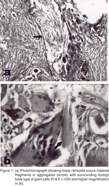
On gross examination, the serosal surface of the resected segment of the jejunum revealed extensive areas of congestion and multiple adhesions. Near one resected margin, a perforated area was seen measuring 3 cm x 1.5 cm and the bowel wall at the margin of the perforation was thickened. The mucosa of the entire length of the bowel was covered with exudate and the mucosal folds were congested, bulky and oedematous. Histopathological examination of the margins of the perforation revealed an ulcerated jejunal mucosa with marked congestion, oedema and inflammation. The submucosa, muscularis propria and serosa showed congestion and dense acute and chronic inflammatory infiltrate. There was marked fibrosis with perivascular and focal collection of lymphoid cell infiltrate. Linear refractile artificial material of suture fragments were seen in aggregates and scattered amidst the inflammatory infiltrate surrounded by numerous foreign body type of giant cells (Figures 1 A and B). Dense acute inflammatory exudate was seen over the serosal aspect. A diagnosis of jejunal perforation with foreign body granulomas, (most likely suture granulomas) was made in view of the previous surgeries.

Discussion
Intra-abdominal foreign body granulomas usually resulting from previous surgery have varied clinical and radiological manifestations. The nuclei of such granulomas can be made up of sutures, talc/starch from surgical gloves, cellulose fibres from surgical gowns, oxidised cellulose from hemostatic agents and mineral oil/paraffin used to prevent adhesions. The peritoneum and mesocolon are commonly involved,[2,3] and either remain asymptomatic or can initiate any type of tissue response. It can lead to aseptic fibrinous reaction resulting in the formation of adhesion, encapsulation and/or foreign body granuloma or an exudative type of tissue reaction resulting in abscess formation.4,5 In either case a mass lesion is formed either in the immediate post-operative period or months or years later.
Foreign body granulomas are more common in patients who have undergone recent surgery, suture granulomas comprising the most important type.[6] The suture material itself and the ischemia caused by tightening of the sutures can initiate an inflammatory reaction and result in adhesion formation. This depends on the rate of resorption of the suture material by the body, the highest rate occurring in the first year post-operatively. This explains why the incidence of suture granulomas is less in cases that have been operated on more than two years before the time of presentation than in cases operated on within a two-year period. Sometimes military peritoneal nodules with serosal inflammation and adhesions can mimic metastatic carcinoma, tuberculosis or Crohn’s disease.[3] Intra-abdominal adhesions are more frequent in patients with multiple prior surgeries as was also seen in this case, and in patients with untreated intra-abdominal inflammatory conditions, previous post-operative intraabdominal complications and where adhesions are already present due to previous laparotomy. [6] The mortality rate is very high and is usually the result of secondary intestinal perforation, peritonitis or sepsis. Malignancy occurring in longstanding cases of foreign body granuloma poses great diagnostic difficulty especially when associated with intraabdominalabscess or granulation tissue.[7]
A solitary foreign body granuloma can be mistaken for a malignant neoplasm unless the foreign body is visualized radiologically. Sometimes the non-absorbable hemostatic material lacks radio-opaque markers, or these are degraded in long-standing cases and may not be visible radiologically.[4] When the clinical course is chronic, the lesion appears hypervascular on angiography. Although the imaging modalities such as ultrasonography, computed tomography and magnetic resonance imaging are useful in detecting foreign body granulomas, a few cases lack the typical radiological findings, and hence remain difficult to diagnose. The presence of sutures is barely visualised grossly or on radiology.
Treatment includes peritoneal cleansing with systemic antibiotics. Simple plication of the perforation is an option or resection and anastomosis where perforations are multiple. In some cases, extended resection with removal of contiguous structures with thorough sampling of the resected specimen may be required for a definitive diagnosis.
The patient in the present case presented with an intraabdominal abscess. Pre-operative diagnosis was not possible as the symptoms were non-specific and radiologically there was no significant finding other than the abscess. The indwelling suture material from the previous surgery must have caused an exudative type of peritoneal reaction resulting in the formation of adhesions, perforation and abscess. Secondary intra-abdominal granulomatous reaction to the suture material was also evident on microscopy.
To conclude, intra-abdominal foreign body granuloma is a rare but potentially serious complication of laparotomy. It may have varied clinical presentation ranging from asymptomatic small intestine perforation and obscure peritonitis to the atypical findings of a malignant neoplasm. Indwelling foreign material is an important cause of intra-abdominal adhesions, suture granulomas being fairly common. An accurate diagnosis via surgical intervention and histopathological examination can avoid suspicion of malignancy and also reduce themortality rate associated with generalised peritonitis. Strategies such as restricting the use of suture material, forgoing closure of the peritoneum and using powder-free gloves may help prevent the occurrence of intra-abdominal foreign body granulomas following laparotomy.[3,8] Careful handling of the tissue may minimise contamination with foreign material so as to avoid complications of intestinal obstruction, infertility and a re-operation.
References
1. Rajagopalan AE, Pickleman J. Free perforation of the small intestine. Ann Surg. 1982;196:576–9.
2. Yamamoto T, Hirohashi K, Iwasaki H, Kubo S, Tanaka Y, Yamasaki K, et al. Pseudotumor of the omentum with a fishbone nucleus. J Gastroenterol Hepatol. 2007;22:597–600.
3. Rosai J. Rosai and Ackerman’s Surgical Pathology. 9th edition, Volume 2, St. Louis, Mosby, 2004, Chapter 26;2373–2415.
4. Yamamura N, Nakajima K, Takahashi T, Uemura M, Nishitani A, Souma Y, et al. Intra-abdominal textiloma. a retained surgical sponge mimicking a gastrointestinal stromal tumor: Report of a case. Surg Today. 2008;38:552–4.
5. Nakajo M, Jinnouchi S, Tateno R, Nakajo M. 18F-FDG PET/CT findings of a right subphrenic foreign body granuloma. Ann Nucl Med. 2006;20:553–6.
6. Luijendijk RW, de Lange DC, Wauters CC, Hop WC, Duron JJ, Pailler JL, et al. Foreign material in postoperative adhesions. Ann Surg. 1996;223:242–8.
7. Joo YT, Jeong CY, Jung EJ, Joo YT, Jeong CY, Jung EJ, et al. Intraabdominal angiosarcoma developing in a capsule of a foreign body: report of a case with associated haemorrhagic diathesis. World J Surg Oncol. 2005;3:60.
8. Luijendijk RW, Wauters CC, Voormolen MH, Hop WC, Jeekel J. Intra-abdominal adhesions and foreign-body granulomas following earlier laparotomy. Ned Tijdschr Geneeskd. [Dutch] 1994;138:717–21.
|
|
|
 |
|
|