48uep6bbphidvals|321
48uep6bbph|2000F98CTab_Articles|Fulltext
There have been few reports on peritoneal loose bodies. Here, we are reporting a rare case of peritoneal loose body in a patient who presented with bleeding per rectum along with rare CT images of loose peritoneal body.
Case report
A 65 yr old male came to our OPD with chief complaints of bleeding per rectum since 1 year and dull aching pain in lower abdomen since 4 months. Patient had bleeding per rectum intermittently, bleeding used to stop spontaneously only to recur after few days. Patient had history of intermittent constipation since 2 years and persistent dull aching pain in infraumbilical region without any relation to food , defecation or micturation and without any aggravating or relieving factors.
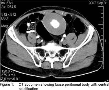
On physical examination, there was intraperitoneal lump in right iliac region about 10x10cm spherical in shape, with well defined margins, smooth surface, firm in consistency and freely mobile. Per rectal examination was normal. Proctoscopic examination revealed grade II haemorrhoids at 3 and 7 o’clock position. CT abdomen (Figure 1) revealed heterogeneously enhancing mass of size 8.5x9.5cm with calcification at center of it, suggestive of duplication cyst dermoid cyst in right iliac fossa ,as reported by radiologist.
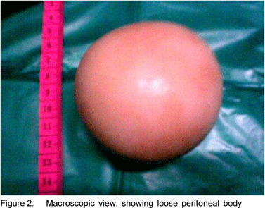
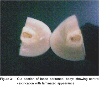
Patient’s Hb was 9 gms, rest other routine investigations (total and differential count, liver function test and urine examination) were within normal limits. On exploratory laparotomy, peritoneal loose body of size 9.5(length) x 8.6(width) cm (Figure 2) found in right iliac region having smooth surface, dirty white in colour and having consistency like boiled egg. Cut surface revealed central calcification with laminated appearance towards periphery (Figure 3). Rest of the abdominal cavity was normal .Post operative recovery was uneventful. Patient’s complaints of bleeding per rectum and constipation were totally relieved postoperatively. Post operative proctoscopic examination after 3 weeks revealed regression of haemorrhoids.


Discussion
Harrigan first described free-lying appendix epiploica.[1] The appendices epiploica are small pouches of peritoneum containing variable amount of fatty tissue.[2]The term loose body implies something which has worked free from the lining of abdomen, resembling the loose bodies found in joints.[3] Hunt (1919) believed that necrosis of pedicle of appendix epiploica was a possible source of loose peritoneal body.[4] Virchow in 1863 proposed the theory of formation of loose peritoneal body.[2] He said that as a result of obesity, the appendices epiploicae increase in fat content and their weight increases, which in turn leads to gradual and progressive obliteration or obstruction of blood vessels of the pedicle. They may undergo torsion, infarction and finally amputation. However, Patterson in 1933 suspected that ischemia as a result of torsion or inflammation is dominant etiologic factor that leads to infarction and amputation.[5] In case of chronic torsion of appendix epiploica, the blood supply is shut off, subsequent saponification and calcification of fat contents occur, the pedicle atrophies. Finally the appendix epiploica gets detached from colon and becomes a loose peritoneal body.[6] Over the years peritoneal reaction to this freely moving appendix epiploica and peritoneal serum deposition over it causes its enlargement. Thus, at center of appendix epiploica there is calcification surrounded by thick laminated fibrinoid material. In present case, the histopathology showed thick laminated hyaline wall with inner layers showing diffuse granular calcification. Takada et al[6] and Nomura et al[7] preoperatively suspected loose peritoneal body as calcified leiomyoma. The loose peritoneal bodies are of variable sizesand number, arise as a result of chronic process. Mohri et al[8] reported a giant loose peritoneal body of size 9.5cm.They mentioned that the body has increased in size from 7.3 cm to 9.5 cm in the span of 5years. Peritoneum reacts by depositing fibrinoid material over it and it goes on increasing in size. Loose peritoneal body may give rise to no definite symptoms or may present with various symptoms like low abdominal pain or constipation[3] or even may present as acute retention of urine.[9] Many times they are found incidentally at laparotomy or in a cadaver.[3]
In the present case, patient had chronic constipation leading to hemorrhoids and these symptoms were relieved after surgery .So, we suspect that the loose peritoneal body may be causing pressure on rectum intermittently. Probably, because of its weight (108gms in present case) and smooth surface it might be getting lodged in pelvis intermittently giving rise to above symptoms.
References
1. Harrigan AH. Torsion and inflammation of the appendix epiploicae. Ann Surg. 1917;66:467–78.
2. Borg SA, Whitehouse GH, Griffiths GJ. A mobile calcified amputed appendix epiploica. Am J Rentogenol. 1976;127:349–50.
3. Ross JA, McQueen A. Peritoneal loose bodies. British J Surg. 1947/48;35:313–7.
4. Hunt VC: Torsion of appendices epiploicae. Ann Surg.
5. 1919;69:31–46.Patterson DC. Appedices epiploicae. N Engl J Med. 1933;209:1255–9.
6. Takada A, Moriya Y, Muramatsu Y, Sagae T. A case of giant peritoneal loose bodies mimicking calcified leiomyoma originating from the rectum. Jpn J Clin Oncol. 1998;28:441–2.
7. Nomura H, Hata F, Yasoshima T, Kuwahara S, Naohara T, Nishimori H, et.al. Giant loose peritoneal body in pelvic cavity:Report of case. Surg Today. 2003;33:791–3.
8. Mohri T, Kato T, Suzuki H. A giant peritoneal loose body: report of a case. Am Surg. 2007;73:895–6.
9. Bhandarwar AH, Desai VV, Gajbhiye RN, Deshraj BP. Acute retention of urine due to a loose peritoneal body. Br J Urol. 1996;78:951–2.