48uep6bbphidvals|593
48uep6bbph|2000F98CTab_Articles|Fulltext
Pancreatic calcification commonly involves the pancreatic duct. Parenchymal calcification and calcification of pseudocysts are uncommon.
Case report
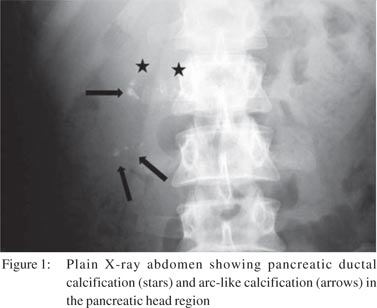
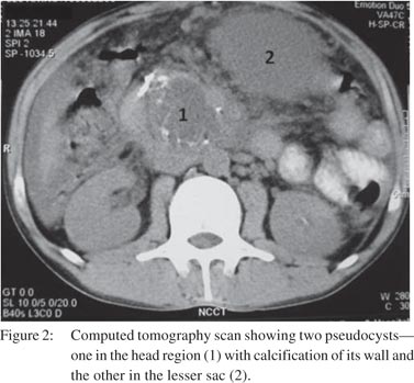
A 35-year-old man was admitted to our hospital with abdominal pain and intermittent vomiting for 15 days. The patient had been consuming 50–100 mL of alcohol for the past 12 years. He was investigated for alcoholic pancreatitis. The serum amylase was 218 IU/mL and lipase 2530 IU/mL. Plain X-ray abdomen showed pancreatic ductal calcification (stars) and arc-like calcification (arrows) in the pancreatic head region (Figure 1). Ultrasound of the abdomen revealed pancreatic ductal calcification and two pseudocysts—one 4.1 × 3.2 × 3.7 cm in the head of the pancreas1 and the other 8.9 × 7.2 × 7.7 cm in the lesser sac.2 The pseudocyst in the pancreatic head showed calcification (arrows) along its wall. Plain and contrast-enhanced computed tomography (CECT) showed a pseudocyst in the head region with calcification of its wall and the other in the lesser sac (Figure 2). Calcification of the pancreatic duct was seen separately . The patient was managed conservatively with enzyme supplements, proton pump inhibitors and anti-oxidants; and his symptoms showed improvement. The lesser sac collection resolved on follow-up but the calcified smaller collection has persisted, though he has been pain-free for over 9 months.


Discussion
Calcification of pancreatic pseudocysts is uncommon. It usually occurs when a pseudocyst escapes the initial diagnosis and calcification ensues with the passage of time.1 Calcification in a pseuodocyst has an egg shell-like appearance and must be differentiated from the nodular and granular calcification of chronic pancreatits. Alcoholic and idiopathic pancreatitis are the two common causes of the chronic pancreatitis. Most often, such calcification is within the pancreatic duct. Sometimes the pancreatic duct can get calcified and pancreatic trauma or infarction with subsequent haemorrhage can lead to parenchymal calcification.[1] Our patient had both, i.e. ductal and pseudocyst calcification. Rowland et al. have also reported the simultaneous occurrence of ductal and pseudocyst calcification.[2]
Other causes of pancreatic calcification are cystic fibrosis, hyperparathyroidism, mucinous cystadenoma, adenocarcinoma and echinococcal cyst of the pancreas.[3] The last three causes can have curvilinear calcification. At times, curvilinear calcification seen on a plain X-ray or CT scan can be the first clue to the underlying chronic pancreatitis. As reported in a case by Munn et al., calcification may be picked up as late as 40 years after the pancreatic trauma.[1]
References
- Munn J, Altergott R, Prinz RA. Calcified pancreatic pseudocysts. Surgery. 1987;101:511–7.
- Rowland R, Stewenson GW, Faris IB. Calcified pancreatic pseudocyst—a report of two cases. Austras Radiol. 1985;29:248–50.
- Ring EJ, Eaton SR, Ferruci JT, Short WF. Differential diagnosis of pancreatic calcification. Am J Roentgenol. 1973;177:446–52.