48uep6bbphidcol2|ID
48uep6bbphidvals|2963
48uep6bbph|2000F98CTab_Articles|Fulltext
Granulosa cell tumors (GCT) constitute 2 to 5% of ovarian malignancies although they are the most common malignant sex cord—stromal tumours of the ovary1,2. They can rarely arise in locations other than the ovary and may be derived from the mesenchyme of the genital ridge.2 The adult subtype usually presents after menopause with per vaginal bleeding and pain. Women who have undergone oophorectomy alsohave the potential to develop GCT2. The primary extraovarian GCT is an extremely rare tumour.3
Case Report
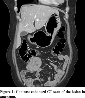
A 55-year-old female presented with continuous dull aching pain over the upper abdomen for the last 4 years. She also had complaints of intermittent nausea. She had previously undergone hysterectomy with bilateral salpingo-oophorectomy elsewhere 12 years earlier for uterine leiomyoma. Biopsy did not reveal anything abnormal in the ovaries. Abdominal examination revealed an ill-defined mass palpable in the umbilical region. On investigation, her blood parameters were within normal limit. Sonography of the abdomen showed a mass in the right hypochondriac region adherent to the small bowel loops. The patient underwent computed tomography (CT) scan abdomen which was suggestive of an omental tumour close to the distal jejunal loop, probably a gastro-intestinal stromal tumour (GIST) (Figure 1). A diagnostic laparoscopy showed an omental mass with areas of haemorrhage close to the distal ileum. The omental tumour was removed in-toto and sent for histopathology.
Gross examination of the mass revealed greyish brown, soft nodular lesion with haemorrhagic areas measuring 7x5 cm. Cut section revealed solid homogenous greyish white tumour with small cystic areas and multiple areas of haemorrhages (Figure 2).
Microscopic findings showed a solid cellular tumour composed of sheets of oval tumor cells with interspersed thin walled blood vessels. The tumour cells had eosinophilic cytoplasm, fairly regular nucle and nuclear grooving with small nucleoli. Few scattered mitotic figures were seen (Figure 3). Based on the classic histopathologic features observed, a diagnosis of granulosa cell tumour made. Immunohistochemistry (IHC) was positive for inhibin and negative for cytokeratin, thus confirming the diagnosis of extra-ovarian granulosa cell tumour (adult type) of the omentum.
Discussion
Granulosa cell tumors (GCT) are malignant sex cord—stromal tumors of the ovary. They constitute 2% of all ovarian tumors.1 They can recur or metastasise many years after initial treatment. Rarely GCTs can develop at extra-ovarian sites. The possibility of metastasis has to be ruled out before making a diagnosis of extraovarian GCT. Extra-ovarian granulosa cell tumour is an unusual tumour with only few cases reported in English literature and rarely reported from the Indian subcontinent.4,6 Extraovarian granulosa cell tumours can develop in the retroperitoneum4,7, broad ligament5, mesentery, omentum, liver, adrenals, and so forth4. The tumour cells are thought to derive their origin from ectopic gonadal stromal tissue from the mesonephros.4 No extra-ovarian GCT has been reported to have originated from omentum.
GCTs vary in their gross appearance. Most are partly cystic and partly solid. Intracystichaemorrhage is common1. Microscopically, the tumour cells resemble normal granulosa cells. They are small with uniform round or oval hyperchromatic nuclei with finely granular chromatin and longitudinal nuclear grooves or fold1. They show microfollicular, macrofollicular, trabecular or diffuse patter1. Similar histologic findings were observed in our case.
Extraovarian GCT should be differentiated from other metastatic carcinomas of the ovary having similar morphology. Inhibin and cytokeratin can help in differentiating these tumours. GCTsare positive for inhibin and negative for cytokeratin. GCTs also have to be differentiated from other tumours such as small cell carcinoma, undifferentiated carcinoma, endometrial stromal sarcoma, carcinoid, and lymphom1. These tumoursare usually inhibin negative. Sometimes, IHC for cytokeratin, Calretinin, Melan-A, FLI-I, CD 99 can help in diagnosing and differentiating these tumours from GCT.
In our case, the patient had a history of hysterectomy, with bilateral salpingo-oophorectomy 12 years ago for leiomyoma with no evidence of GCT of the ovary in the previous histopathology report. The patient presented with mass in the abdomen and pain due to contained tumour rupture, which is a well-known complication of GCT. Naniwadekar MR et al also described similar tumor in mesentery with prior hysterectomy and bilateral oophorectomy. In our case, hormonal studies were not done, as the diagnosis of GCT was not suspected. The histopathology features of the present tumourwere typical of GCT with pale & round to oval granulosa cells with diffuse histological pattern and characteristic “coffee-bean” nuclei. The tumourwas inhibin positive and cytokeratin negative, thus confirming the diagnosis of GCT.
Conclusion
GCTs can arise in multiple locations other than the ovary and are considered to be derived from the mesenchyme of the genital ridge. Although rare, women who have undergone oophorectomy may also develop GCT at an extraovarian location includingthe omentum. A diagnosis of extraovarian GCT can be made by excluding any previous history of GCT of either ovary. Immunohistochemistry may help in differentiating GCT from other neoplasms.
References
- Z. Charles and W. N. Brenda, “The ovary and fallopian tube,” in Silverberg’s Principles and Practice of Surgical Pathology and Cyopathology, S. G. Silverberg, Ed., vol. 2, pp. 2015–2017, Churchill Livingstone Elsevier, St. Louis, Miss, USA, 4th edition, 2006.
- Chou CF, Huang WC. Granulosa cell tumor of the ovary. Tzu-Chi Medical Journal. 2016 ;28:84.
- Kim SH, Park HJ, Linton JA, Shin DH, Yang WI, Chung WY, Kim YT. Extraovarian granulosa cell tumor. Yonsei Medical Journal. 2001;42:360-3.
- Paul PC, Chakraborty J, Chakrabarti S, Chattopadhyay B. Extraovarian granulosa cell tumor. Indian Journal of Pathology and Microbiology. 2009 Apr 1;52(2):231.
- Sakai Y. Granulosa cell tumor arising in the wall of müllerian cyst of the broad ligament: report of a case and immunohistochemical study. Archives of gynecology and obstetrics. 2007 ;275:145-8.
- Naniwadekar MR, Patil NJ. Extraovarian granulosa cell tumor of mesentery: A case report. Pathology research international. 2010;2010.
- Keitoku M, Konishi I, Nanbu K, Yamamoto S, Mandai M, Kataoka N, Oishi T, Mori T. Extraovarian sex cord-stromal tumor: case report and review of the literature. International journal of gynecological pathology: official journal of the International Society of Gynecological Pathologists. 1997;16:180-5.