48uep6bbphidvals|215
48uep6bbphidcol2|ID
48uep6bbph|2000F98CTab_Articles|Fulltext
Benign tumours account for less than 3% of all esophageal neoplasms and are classified according to whether they are intramural or intraluminal. Intraluminal esophageal neoplasms are extremely rare and often lead to diagnostic dilemma. We report the case of a patient with giant fibrovascular polyp (FVP) with illustrative images, and review the literature on the subject.
Case Report
A 60-year-old male patient presented with the complaint of gradually progressive dysphagia for solid food of six months duration. He also gave history of regurgitation of food and occasional choking sensation when asleep. Clinical examination did not reveal any abnormality except for anaemia, with a hemoglobin level of 6.8 g/dL. His chest radiograph revealed a right paratracheal opacity with well-defined lateral margins. The vocal cords were normal on indirect laryngoscopy. Fibre-optic upper gastrointestinal endoscopy revealed multiple compressible soft polypoidal intraluminal projections with smooth, whitish overlying mucosa. The mass extended throughout the esophagus and projected into the stomach through the gastro-esophageal junction.
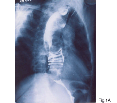
On barium swallow study, (Figure 1A) a dilated esophagus with lobulated margins in its entire extent producing a ‘haustrated’ appearance similar to that of the colon was seen. There were multiple intraluminal filling defects with smooth margins noted within the dilated esophageal lumen. However, there was no obstruction to the flow of ingested barium suspension through the esophagus despite the presence of the multiple filling defects.


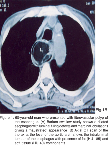
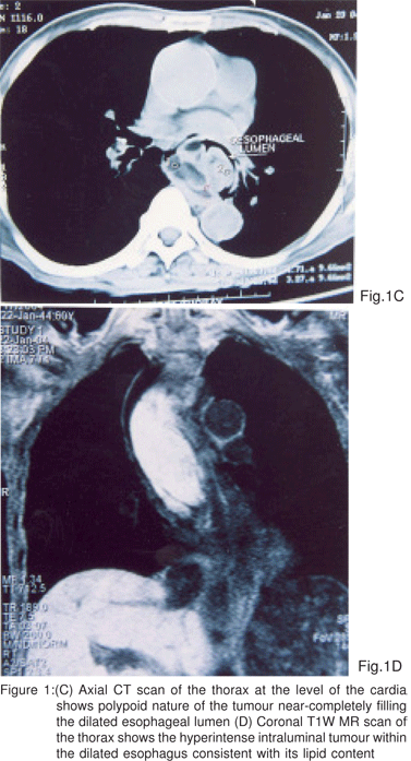
Contrast enhanced computerised tomography (CT) of the thorax (Figures 1B & C) demonstrated the presence of multiple smooth marginated polypoidal filling defects all through the esophageal lumen starting just distal to the cricopharynx and reaching inferiorly to the gastro-esophageal junction, without any wall thickening or invasion. The polyps consisted of predominantly fatty tissue (HU values 62–85), with areas ofsoft tissue density (HU value 35-40) and few focal calcifications. Multiple small vascular channels were seen coursing through the mass on the post-contrast study. We arrived at a provisional diagnosis of fibro-vascular polyp of the esophagus based on the clinical presentation and imaging findings.
An MRI study of the thorax delineated the entire extent of the polypoidal lesion, and the lesion was best illustrated in the coronal T1-weighted images, which also demonstrated the predominant hyperintense fatty content of the polyp (Figure 1D).

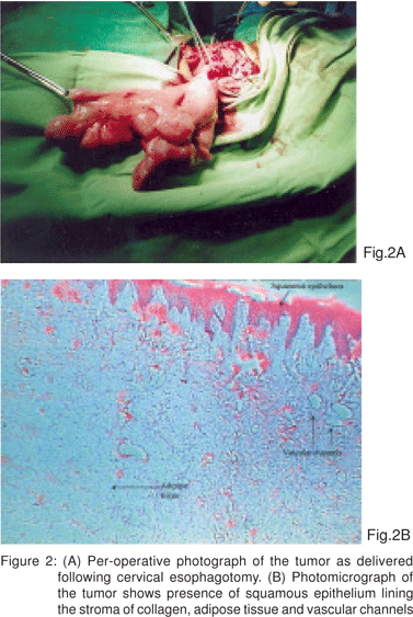
The patient underwent cervical esophagotomy and surgical extirpation of the entire polypoidal mass (Figure 2A), which measured approximately 19 cm in length, and had an uneventful post-operative recovery. Histology of the polyp revealed that the mass was composed of a mixture of adipose and fibrovascular tissue, lined by normal squamous epithelium (Figure 2B), confirming the diagnosis of fibrovascular polyp (FVP).
On six-monthly follow up over the past 5 years, the patient is well and symptom-free and upper gastrointestinal endoscopy has not shown any local recurrence.

Discussion
Benign tumours of the esophagus are rare,[1,2,3] the majority comprise intramural tumours, which may be multiple and usually arise from the esophageal musculature in its middle and distal thirds; the commonest variety is the leiomyoma. Intraluminal lesions on the other hand are of mucosal origin, solitary in nature and generally arise from the proximal third of the esophagus. Fibro-vascular polyp (FVP) is the commonest intraluminal tumour of the esophagus.2 A Pubmed search revealed only about 40 cases reported in world literature in the last 30 years, and only a single case series of 16 fibrovascular polyps has been compiled, at the Armed Forces Institute of Pathology.[1]
FVPs are more common in the male patients, with peak incidence in the 6th and 7th decades. [1,2]However, cases occurring in children or infants[4,5] and in women[6] have also been reported. As the name implies, these are composed of fibrous tissue, adipose tissue and vascular structures, and are usually covered by normal esophageal squamous epithelium. The tissue composition and morphologic appearances of the FVP combine to award them multiple synonyms, such as pedunculated lipoma, fibrolipoma, fibroepithelial polyp, myxofibroma and angiofibrolipoma.[3]
The site of origin of the FVP within the oesophagus is thought to be from the Laimer-Haeckemann area (or Laimer’s triangle), which is a region of relative muscular deficiency just inferior to the cricopharyngeus.[7] The FVP originates as nodular submucosal thickening and progressively elongates under the combined downward traction effects of oesophageal peristalsis and pressure of the passing food material.[1,8] FVPs of giant proportions, including those more than 20 cm in length have been reported.[2]
Patients are usually asymptomatic in the early stages of the disease. Most patients have lesions exceeding 7 cm in length at the time of presentation.[2,9] Chronic non-specific symptoms such as retrosternal discomfort, chest heaviness and loss of weight are common, and onset of dysphagia points towards oesophageal obstruction. Chronic respiratory symptoms may occur due to aspiration as well as pressure effect on the adjacent trachea. Rarely mucosal ulceration may lead to occult gastrointestinal bleeding.[2] The polypoidal pedunculated mass may pass retrograde into the hypopharynx and obstruct the laryngeal inlet.[7] In the series of Levine et al,[1] all 16 patients with giant fibrovascular polyps were symptomatic; dysphagia was the most common complaint (present in 87%), followed by respiratory symptoms (25%) and regurgitation of the polyp into the pharynx or mouth (12%). Although that series did not show examples of asphyxiation, this is a feared complication that can result in sudden death from impingement of the mass onto the larynx. Two patients with giant fibrovascular polyps reported by Owens et al,[7] presented emergently with intermittent airway obstruction following episodes of coughing; the obstruction was relieved by the patients’ clenching of the regurgitated mass between their teeth. Our patient complained of episodes of intermittent airway obstruction that occurred during sleep.
The conventional chest skiagrams usually depict mediastinal widening and some show anterior ‘bowing’ of the trachea.[2] Endoscopy usually reveals a soft, polypoidal, compressible mass covered with intact normal oesophageal mucosa. Barium swallow study delineates multiple intraluminal filling defects, which despite their size do not obstruct the column of barium. ‘Forking’ of the column of contrast at the origin of the FVP is considered a characteristic finding.[1]
CT is the preferred modality for delineation of the intraluminal location of the tumour mass, and objective demonstration of the vascular and lipid contents of the FVP.[8] The presence of fat within an intraluminal esophageal mass lesion is considered diagnostic of FVP. Coronal and sagittal image reconstructions will depict the entire longitudinal extent of the tumour accurately. MRI thorax with T1- weighted coronal images is also diagnostic of FVP and offers a multiplanar imaging advantage, as compared to CT.[6,8]
The treatment is surgical, and the approaches described are – trans-oral, trans-thoracic and trans-cervical.[2] The preferred approach has always been trans-cervical, which accesses the stalk of the polyp, which almost invariably is in the post-cricoid portion of the cervical esophagus.[3] The stalk of the polyp is ligated and divided, and the entire polyp delivered into the neck by traction. Recurrence of the tumour following surgery is rare.9 Endoscopic snare polypectomy10 and endoscopic laser therapy[8,11] are suggested as alternative methods, but are not favoured due to inadequate control of haemorrhage.
Awareness and an accurate pre-operative diagnosis of fibrovascular polyp, averts the morbidity of a needless thoracotomy, for this rare benign condition.
References
1. Levine MS, Buck JL, Pantongrag-Brown L, Buetow PC, Hallman JR, Sobin LH. Fibrovascular polyps of the esophagus: clinical, radiographic, and pathologic findings in 16 patients. AJR Am J Roentgenol. 1996;166:781–7.
2. Belafsky P, Amedee R, Zimmerman J. Giant fibrovascular polyp of the esophagus. South Med J. 1999;92:428–31.
3. Fries MR, Galindo RL, Flint PW, Abraham SC. Giant fibrovascular polyp of the esophagus. Arch Pathol Lab Med. 2002;127:485–7.
4. Paik HC, Han JW, Jung EK, Bae KM, Lee YH. Fibrovascular polyp of the esophagus in an infant. Yonsei Med J. 2001;42:264–6.
5. Seshul MJ, Wiatrak BJ, Galliani CA, Odrezin GT. Pharyngeal fibrovascular polyp in a child. Ann Otol Rhinol Laryngol. 1998;107:797–800.
6. Ascenti G, Racchiusa S, Mazziotti S, Bottari M, Scribano E. Giant fibrovascular polyp of the esophagus: CT and MR findings. Abdom Imaging. 1999;24:109–10.
7. Owens JJ, Donovan DT, Alford EL, McKechnie JC, Franklin DJ, Stewart MG, et al. Life-threatening presentations of fibrovascular esophageal and hypopharyngeal polyps. Ann Otol Rhinol Laryngol. 1994;103:838–42
8. Borges A, Bikhazi H, Wensel JP. Giant fibrovascular polyp of the oropharynx. AJNR Am J Neuroradiol. 1999;20:1979–82.
9. Behar PM, Arena S, Marrangoni AG. Recurrent fibrovascular polyp of the esophagus. Am J Otolaryngol. 1995;16:209–12.
10. Wu MH, Chuang CM, Tseng YL. Giant intraluminal fibrovascular polyp of the esophagus. Hepatogastroenterology. 1998;45:2115–6.
11. Naveau S, Bedossa P, Mallet L, Turner L, Poynard T, Andréani T, et al. Successful ablation of a large fibrovascular polyp of the esophagus by endoscopic Nd:YAG laser therapy. Gastrointest Endosc. 1989; 35:254–6.