K Sambasivaiah, S. Samson, Ramakrishna, V. Prasanna, A Hari Shankar
Narayana Medical College Hospital,
Nellore, Andhra Pradesh, India
Corresponding Author:
Dr. K.Sambasivaiah
Email: sambasvims@yahoo.co.in
Abstract
The common presentation of carcinoma stomach includes haematemesis, malaena and gastric outlet bstruction. Symptoms due to metastases to the liver and lung as part of the disease progression are usually preceded by the detection of a primary in the stomach. Carcinoma stomach, where the primary is silent, and which presents with truncal ataxia and features of hydrocephalus due to isolated metastatic deposit in the cerebellar vermis is exceptionally rare. The prognosis of such patients is poor and the treatment is palliative.
|
48uep6bbphidvals|229 48uep6bbphidcol2|ID 48uep6bbph|2000F98CTab_Articles|Fulltext The presenting features of carcinoma stomach arise due to the location of the primary tumour, and include haematemesis; malaena and gastric outlet obstruction. Carcinoma stomach usually metastasises to the liver and lung and as the disease progresses; rarely metastases to the brain may also occur. However, carcinoma stomach with initial presentation of truncal ataxia and hydrocephalus, as a result of metastasis to the cerebellar vermis is exceptionally rare. The following is a description of just such a case.
Case Report
A forty-one year old male patient presented to our emergency service with complaints of sudden onset, holocranial headache associated with projectile vomiting of 3 weeks duration. Simultaneously he also had difficulty in walking and in talking. His bowel and bladder functions were normal. There was no previous history of ear discharge, exposure to dyes or toxins. There was no history of hypertension, diabetes mellitus, or pulmonary tuberculosis. He was a smoker and onsumed alcohol regularly.
On clinical examination the only finding was that of pallor, everything else was normal. On neurological examination, the higher mental functions were normal. There was dysarthria, and bilateral horizontal gaze evoked nystagmus, and bilateral papilloedema on fundus examination. Motor system examination consisting of tone, power and reflexes were normal and plantars showed flexor response. The sensory examination was normal. The coordination was normal in all four limbs. There was truncal ataxia and an inability to perform tandem gait. No signs of meningeal irritation were present. The skull and spine were normal. Cardiovascular, respiratory and abdominal examinations were normal.
MRI brain with gadolinium contrast revealed a midline vermian mass (Figure 1) measuring 3.6 cm x 2.3 cm x 3.2 cm, with perilesional oedema, and causing a mass effect over the 4th ventricle with resultant obstructive hydrocephalus. The haemoglobin was 10.0 g/dl, and the total white cell count was 12400 cells/ mm3 with polymorphs, lymphocytes, eosinophils and monocytes as 58%, 21%, 14% and 7% respectively, and the erythrocyte sedimentation rate was 10 mm/hr. the random blood sugar was 120 mg/dl, the blood urea was 32 mg/dl, the serum creatinine was 1 mg/dl, the serum sodium was 135 meq/l, the serum potassium was 4.7 meq/l and the serum chloride was 98 meq/l.
Routine urine analysis was normal. Chest x-ray was normal. Ultrasound abdomen was normal.
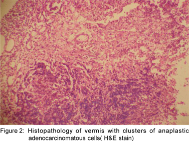
The patient underwent midline sub-occipital craniectomy with excision of the mass. At surgery, there was a well-defined and firm mass in the vermis, which was totally excised. The histopathology depicted the tissue structure of the vermis with clusters of anaplastic adenocarcinomatous cells arranged in sheaths and groups with areas of necrosis and reactive gliosis (Figure 2). The patient’s neurological status improved postoperatively.
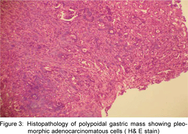
Tumour markers as part of a search for the primary were tested for and included the prostate specific antigen and the carcinoembryonic antigen which were normal. Gastroscopy revealed an exophytic, ulceroproliferative growth in the body and antrum, which on histological examination depicted pleomorphic adenocarcinomatous cells (Figure 3).


Discussion
Carcinoma of the stomach presents with well-known symptoms and signs related to blood loss from the tumour mass or to gastric outlet obstruction. Most commonly stomach cancer spreads contiguously to the perigastric tissues and adjacent organs such as the pancreas and colon; by the lymphatics to the abdominal lymph nodes and haematogenously to the distant organs such as the liver and lung. However, gastric cancer presenting with only atypical features such as truncal ataxia and hydrocephalus resulting from metastasis to the vermis of the cerebellum is exceptionally rare.
One case report of cerebellar metastases due to gastric cancer has been described previously.[1] Few other case reports of gastric cancer with atypical features resulting from metastases to the pineal gland[2,3] and erebellopontine angle[4] have also been described. Often these metastases develop several years after the treatment for carcinoma stomach and metastases to other viscera. The present case is unique, as the primary in the stomach was silent and the patient presented with truncal ataxia and signs of hydrocephalus; further, the liver and the lung, the most frequent sites of visceral metastases for gastric cancer were spared. The molecular mechanisms responsible for exclusive intracranial metastases are not known.The treatment for intracranial metastases secondary to carcinoma stomach is usually palliative, as the prognosis of these patients is poor. Whole brain radiotherapy may be useful in symptom regression; the impact of systemic chemotherapy on survival is not known.
References
1. Perri F, Bisceglia M, Giannatempo GM, Andriulli A. Cerebellar metastasis as a unique presenting feature of gastric cancer. J Clin Gastroenterol. 2001;33:80–1.
2. Kanai H, Yamada K, Aihara N, Watanabe K. Pineal region metastasis appearing as hypointensity on T2-weighted magnetic resonance imaging—case report. Neurol Med Chir (Tokyo). 2000;40:283–6.
3. Boscherini D, Pintucci M, Mazzucchelli L, Renella R, Pesce G. Neuroendoscopic management of a solitary pineal region tumor. Case report of an adenocarcinoma metastasis. Minim Invasive Neurosurg. 2006;49:247–50.
4. Bono P, Bartoloni Saint Omer F..A rare case of bilateral metastasis in the cerebellopontine angle. Pathologica. 1994;86:324–8.
|