A. Mathew, V. Abraham, U. Shankar, S.V. Thomas, M.R. Jesudason, B. Perakath
Department of Colorectal Surgery,
CMCH,
Vellore, India
Corresponding Author:
Benjamin Perakath
Email- benjamin@cmcvellore.ac.in
Abstract
Tailgut cysts, also called benign retrorectal hamartomas, are uncommon developmental cysts found behind the rectum. Here, we present a rare case of a tailgut cyst associated with uterine anomaly, sacral and vertebral anomalies and vascular duplication, in a young lady who presented with constipation and infertility.
|
48uep6bbphidvals|230 48uep6bbphidcol2|ID 48uep6bbph|2000F98CTab_Articles|Fulltext Tailgut cysts are uncommon retrorectal cystic tumours. We present a rare case of tailgut cyst with subdural extension and associated uterine didelphys, hemisacral deficiency and double inferior vena cava.
Case Report
A 24-year old lady presented with a history of low back pain and difficulty in defecation worsening over the previous 5 years. She was also infertile.
Digital rectal examination revealed a large mass pushing the rectum anteriorly; the overlying rectal mucosa was mobile. The upper border of the mass was not palpable, nor was it palpable per abdomen.
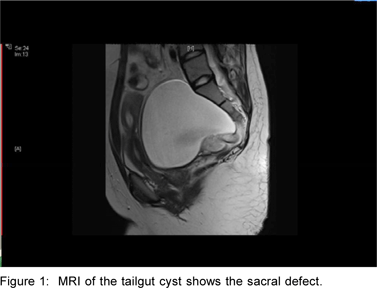
Magnetic resonance imaging of the abdomen and pelvis revealed a cystic mass in the presacral and precoccygeal region measuring 11 cm x 10 cm x 10 cm with solid components. The lower part of the left hemisacrum was deficient with part of the cyst extending into the sacral spinal canal. She also had uterus didelphys with double inferior vena cava and coronal clefts in the 4th and 5th lumbar vertebrae (Figure 1)
Biochemistry, including serum beta human chorionic gonadotropin and alpha-fetoprotein, were normal.
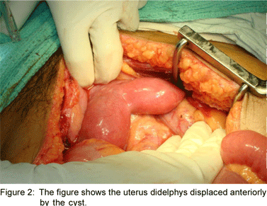
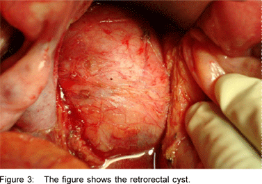
She underwent excision of the retrorectal cyst including its sacral extension (Figures 2 and 3). Peroperatively, there was a cerebrospinal fluid leak which was managed with a lumbar subdural catheter.
Gross examination of the specimen showed an ovoid cystic mass measuring 10 cm x 6 cm x 6cm with a weight of 150 g. The cut section revealed multiple loculations with many smooth walled cysts containing pultaceous material. A focal nodule was seen, which was also cystic on sectioning. The microscopic appearance of the cyst lining was mucin secreting columnar epithelium with adjacent smooth muscle bundles and fibrous connective tissue on the external aspect. The cyst within the nodule was lined by stratified squammous epithelium enclosing lamellated keratin and a focus of granuloma composed of epithelioid cells and foreign body multinucleate giant cells. No immature elements were present.

Discussion
Tailgut cysts (also known as benign retrorectal harmartomas or mucin-secreting cysts) are enteric developmental cysts of the presacral space. Others in this category are the epidermoid, dermoid, cystic rectal duplication and neurenteric cysts.[1]They are found in the female adult population and can be asymptomatic or present with compression symptoms (as in our case) or as pilonidal sinuses, anorectal fistulae or recurrent retrorectal abscesses. Rare case reports of malignant transformation[2] and carcinoid tumour[3] arising in a tailgut cyst have been reported.
Whereas several cases of tailgut cysts per se, as well as those with sacral defects have been reported[2], their association with uterine didelphys, vertebral anomalies and double IVC have not been reported. In 1981, Currarino[4] described a syndrome in infants which consists of a triad of presacral cystic masses, sacral agenesis and anorectal malformation. These have an autosomal dominant inheritance associated with mutations in the HLXB9 gene.[5] These presacral masses comprised the anterior sacral meningocele, cystic teratomas or enteric cysts.
A literature search revealed that rectal duplication cysts[6] and anterior sacral meningoceles[1] have been associated with uterus didelphys but only one other case report of a tailgut cyst associated with uterine didelphys was found. This was the case of a twelve-year old girl who had mental retardation, and had hypothyroidism, sacral and coccygeal agenesis but no chromosomal anomalies.[7]
Abnormalities of the lower urinary and genital tracts as a result of Mullerian and Wollfian duct abnormalities have been reported but none with retrorectal cysts viz. the Mayer Rokitansky syndrome, which includes uterine didelphys, imperforate vagina and renal agenesis.[8]


Double inferior vena cava in itself is a rarity. There are isolated case reports of its association with renal aplasia[9], congenital hepatic fibrosis and lung dysgenesis. No report of double IVC with retrorectal cysts has been published till date.
Embryologically, tailgut cysts are thought to be the persistent part of the hind gut in the region of the embryonic tail that normally involutes.[1,2] Pathologically, tailgut cysts are usually multicystic or multiloculated and lined by a variety of epithelia (stratified squammous, transitional, stratified columnar, mucinous or ciliated columnar, ciliated pseudostratified columnar or gastric). Well formed but disorganised smooth muscle fibres are focally present in its wall unlike the well formed continuous two layer muscle coat seen in rectal duplication cysts. They are dissimilar to benign cystic teratomas which contain distinct dermal appendages, neural elements or mesenchymal derivatives like cartilage or bone.[1]
Excision of the tailgut cyst results in its cure. This case was reported to highlight the congenital anomalies that can also be associated with this condition.
References
1. Dahan H, Arrivé L, Wendum D, Docou le Pointe H, Djouhri H, Tubiana JM. Retrorectal developmental cysts in adults: clinical and radiologic-histopathologic review, differential diagnosis, and treatment. Radiographics. 2001;21:575–84.
2. Prasad AR, Amin MB, Randolph TL, Lee CS, Ma CK. Retrorectal cystic hamartoma: report of 5 cases With Malignancy Arising in 2. Arch Pathol Lab Med. 2000;24:725–9.
3. Horenstein MG, Erlandson RA, Gonzalez-Cueto DM, Rosai J. Presacral carcinoid tumors: a report of 3 cases and review of literature. Am J Pathol. 1998;22:251–5.
4. Currarino G, Coln D, Votteler T. Triad of anorectal, sacral and presacral anomlies. AJR Am J Roentgenol. 1981;137:395–8.
5. Kim IS, Oh SY, Choi SJ, Kim JH, Park KH, Park HK, et al. Clinical and genetic analysis of HLXB9 gene in Korean patients with Currarino syndrome. J Hum Genet. 2007;52:698–701.
6. Nour S, Kumar D, Dickson JA. Anorectal malformations with sacral bony abnormalities. Arch Dis Child. 1989;64:1618–20.
7. Galluzzo ML, Bailez MM, Reusmann A, Gonzalez R, Davila MT. Tailgut cyst (Retrorectal hamartoma): Report of a pediatric case. Pediatr Dev Pathol 2007;22:1.
8. Stassart JP, Nagel TC, Prem KA, Phipps WR. Uterus didelphys, obstructed hemivagina and ipsilateral renal agenesis: the University of Minnesota experience. Fertil Steril. 1992; 57:756–61.
9. Gayer G, Zissin R Strauss S, Hertz M. IVC anomalies and right renal aplasia on CT: a possible link? Abdom Imaging. 2003;28:395–9.
|