48uep6bbphidvals|254
48uep6bbphidcol4|ID
48uep6bbph|2000F98CTab_Articles|Fulltext
Salmonella is a gram-negative bacilli of the Enterobacteriaceae family, with approximately 2000 known serotypes and variants. Non typhoidal salmonellosis is the disease called by any serotype except S. Typhi and S. Paratyphi1. The incidence of non-typhoidal salmonella has increased in the last 2 decades in Europe and North America2, and it is estimated that 1.4 million cases occur annually in the United States resulting in 15,000 hospitalizations and 400 deaths3. Salmonella Enteritidis and S. Typhimurium are the most frequently isolated serotypes responsible for the infections2. Computed Tomography (CT) findings of Salmonella colitis have rarely been reported in the published work, despite the high incidence of non typhoidal salmonellosis in the general population. We herein, present a rare case of Salmonella pancolitis in a non immunocompromized young male patient who presented with sepsis and acute abdomen and the CT findings which indicated acute colitis thus averting an unnecessary laparotomy. A review of the literature has also been done.
Methods
A 21- year- old male with an unremarkable medical history presented with a 24-hour history of high fever (39,8-40,40C), 4 episodes of watery diarrhea and severe abdominal pain. Although the pain was initially periumbilical, it progressively became worse and generalized.
Upon admission, physical examination showed a distressed patient with temperature of 40.20C, respiratory rate of 17 breaths/min, blood pressure of 95/55 mm Hg, and pulse rate of 120 beats/min.Abdominal examination revealed diffuse tenderness, guarding and rebound tenderness, whereas bowel sounds were absent.
A haemogram revealed a white cell count of 11,200/ mm3 with 76.2% neutrophils, and a haematocrit of 36.4%. Liver function tests and biochemical markers were all within normalranges, except C-reactive protein which was markedly elevated (185 U; range 0-5). In addition, coagulation studies revealed a prolonged prothrombin time (PT) of 15.1sec (range 9-13), international normalized ratio (INR) 1.27 (range 0.85-1.15) and activated partial thromboplastin time (APTT) 41.1 sec (range 26-38).
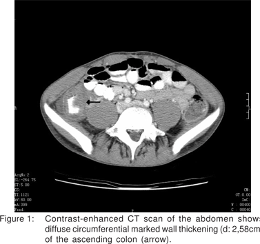
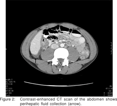
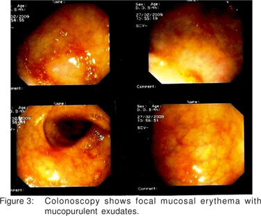
Abdominal ultrasonography revealed free intraperitoneal fluid collection whereas chest and abdominal radiographs were unremarkable. Due to acute abdominal findings and signs of sepsis, an urgent abdominal CT scan was obtained which revealed marked thickening mainly involving the right colon wall (Figure 1), along with ascites (Figure 2). Based on these findings the diagnosis of acute infectious colitis was considered and thus we decided not to proceed to an exploratory laparotomy. After blood and stool cultures were obtained, replacement therapy with fluids and electrolytes was initiated and the patient was placed on antibacterial therapy with ciprofloxacin and metronidazole. After 12 hours, his general condition began to improve steadily and he was discharged home 6 days later, after complete resolution of his symptoms. Stool cultures yielded Salmonella spp, whereas blood cultures were negative. Colonoscopy performed after the acute phase of the disease revealed findings consistent with infectious colitis (Figure.3).
Discussion
Salmonella gastroenteritis is the most common clinical syndrome caused by non-typhoidal salmonella. After an incubation period of 6-48h patients usually present with nausea, vomiting, abdominal cramps and diarrhea[1]. Invasive disease with bacteremia has been reported in fewer than 5% of the cases4, whereas unnecessary surgical interventions to treat suspected acute abdominal conditions have been performed in patients with salmonellosis, increasing morbidity and mortality rates[2]. Clinical presentation is often indistinguishable from gastroenteritis caused by other pathogens. However, most patients have mild to moderate symptoms and the disease tends to run a short self-limited course[5]. In our case, although the patient was nonimmunocompromized he had a very severe clinical course. We speculate that this was likely due to the ingested dose of the bacteria as it has been identified as an important determinant in the incubation period, symptoms and severity of acute salmonellosis[6].
Colonic involvement in salmonellosis was first reported in 1969 when Boyd[7] described necropsy findings of 6 patients with infections caused by S. typhimurium. The clinical course of salmonella colitis varies from 1 week to 2- 3 months with an average duration of 3 weeks[1]. Early diagnosis and appropriate treatment is mandatory to prevent potentially serious complications including toxic megacolon requiring aggressive therapy, sepsis and bleeding[1,8].
Despite the high incidence of salmonellosis in the general population there are only a few reports in the literature describing CT or other radiological findings of salmonella colitis[5,9,10,11,12]. This likely happens because most infected patients do not seek medical assistance, do not undergo diagnostic evaluation and are treated empirically with supportive therapy and broad-spectrum antibiotics[5].
Colonic wall thickness is the main feature on CT indicative of acute colitis. When the lumen of the bowel is distended, the normal thickness is 1-2mm, but when it is collapsed the normal thickness is 3-4mm.[13] Philpotts et al[9] reported a colon wall thickness of 8.0+2.47mm in 11 patients with infectious colitis, whereas in 36% of the patients the findings were exclusively involving the right colon. In another study Horiki et al[10] reported that the thickness of the ascending colon wallwas greater of 10mm in 45% of the patients with salmonella colitis. Finally, Balthazar et al5 reported a slight (3-5mm) circumferential wall thickening of cecum and descending colon in 3 patients with Salmonella infection. Compared to the above reported data, the thickness of the ascending colon wall in our patient was excessive.
The presence of ascites is another finding on CT suggestive of acute colitis and has been reported in 23-46% of the cases[9,10]. CT is mainly used to evaluate patients with more severe clinical presentation and to exclude other abdominal inflammatory processes[5,10].
In most cases the definitive diagnosis of the type of colitis is based on clinical and laboratory data and colonoscopic and biopsy findings[13]. CT findings of colitis are non specific, but some features can be helpful in suggesting a specific diagnosis[9,10], as in our patient. These features may include the amount of wall thickness, extent of the disease, pericolic reaction, ascites, fistulae, and presence of complications[13].
We present a rare case of salmonella pancolitis in a nonimmunocompromized young male patient who presented with sepsis and typical manifestations of acute abdomen. Although definitive diagnosis is made with the isolation of Salmonella spp from stool cultures, however, non specific findings on CT scan should alert the emergency physician consider the diagnosis of acute colitis, in patients presenting with a severe form of the disease, avoiding therefore an unnecessary laparotomy and subsequent morbidity.
References
1. Giannella RA. Infectious enteritis and proctocolitis and bacterial food poisoning. In: Feldman M, Friedman LS, Sleisenger MH, editors. Sleisenger & Fordtran’s Gastrointestinal and Liver Disease.8th ed. Philadelphia, PA: Saunders Elsevier; 2006: p.2333–91.
2. Fisker N, Vinding K, Mølbak K, Hornstrup MK. Clinical review of nontyphoid Salmonella infections from 1991 to 1999 in a Danish county. Clin Infect Dis. 2003;37:e47–52.
3. Voetsch AC, Van Gilder TJ, Angulo FJ, Farley MM, Shallow S, Marcus R, et al. FoodNet estimate of the burden of illness caused by nontyphoidal Salmonella infections in the United States. Clin Infect Dis. 2004;38 Suppl 3:S127–34.
4. Helms M, Simonsen J, Molbak K. Quinolone resistance is associated with increased risk of invasive illness or death during infection with Salmonella serotype Typhimurium. J Infect Dis. 2004;190:1652– 4.
5. Balthazar EJ, Charles HW, Megibow AJ. Salmonella- and Shigellainduced ileitis: CT findings in four patients. J Comput Assist Tomogr. 1996;20:375–8.
6. Mintz ED, Cartter ML, Hadler JL, Wassell JT, Zingeser JA, Tauxe RV. Dose-response effects in an outbreak of Salmonella enteritidis. Epidemiol Infect. 1994;112:13–23.
7. Boyd JF. Salmonella typhimurium, colitis, and pancreatitis. Lancet. 1969;2:901–2.
8. Chaudhuri A, Bekdash BA. Toxic megacolon due to Salmonella: a case report and review of the literature. Int J Colorectal Dis. 2002;17:275–9.
9. Philpotts LE, Heiken JP, Westcott MA, Gore RM. Colitis: use of CT findings in differential diagnosis. Radiology. 1994;190:445–9.
10. Horiki N, Maruyama M, Fujita Y, Suzuki Y, Tanaka T, Imoto I, et al. CT evaluation of infectious colitis. Nippon Shokakibyo Gakkai Zasshi. 2002;99:925–34. Article in Japanese.
11. Ho AC, Horton KM, Curry CA, Schuberth KC, Fishman EK. Radiological Case Review. Appl Radiol. 2002;31:34–36.
12. Nakamura S, Iida M, Tominaga M, Yao T, Hirata N, Fujishima M. Salmonella colitis: assessment with double-contrast barium enema examination in seven patients. Radiology. 1992;184:537–40.
13. Thoeni RF, Cel lo JP. CT imaging of col i t is. Radiology. 2006;240:623–38.