48uep6bbphidvals|323
48uep6bbph|2000F98CTab_Articles|Fulltext
Case 1
Left sided pneumoperitoneum masquerading as gastric bubble
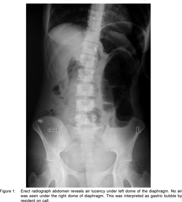
A 19-year-old female presented with history of recurrent colicky pain abdomen, non bilious vomiting, anorexia, and weight loss. She had earlier been investigated in a private institution and was diagnosed as Celiac disease, on the basis of positive IgA anti tissue transglutaminase levels and duodenal biopsy demonstrating altered C:V ratio and increased intraepithelial lymphocytes. There was no clinical response on gluten free diet although her repeat biopsy had shown reversal of changes. At our institution she underwent CT Enteroclysis which demonstrated grossly dilated stomach (reaching the left iliac fossa), first and second part of duodenum, with a stricture in the third part of duodenum. Her Mantoux test was 18 x 18 mm, and on basis of above findings she was started on antitubercular drugs. She underwent balloon dilatation of the stricture, following which she had pain abdomen with distension and had guarding and rigidity, on examination. She was passing faeces and flatus. Radiograph abdomen done after the procedure demonstrated air lucency under left dome of the diaphragm. No air was seen under the right dome of diaphragm. This was interpreted as gastric bubble by resident on call (Figure 1). CT done after 4 hours, because of high clinical suspicion, revealed pneumoperitoneum in the left subphrenic space (Figure 2). The patient was taken for emergency laparotomy which revealed perforation in proximal jejunum and pus in the peritoneal cavity.


Case 2
Giant Exophytic Left Lobe Liver Hemangioma draping the spleen
A 37-year-old female presented to the hospital with incidentally detected mass lesion in the left subphrenic location abutting the borders of the spleen. She had past history of jaundice 6 month ago. Multiphase contrast CT and MR scans were done which revealed a large mass lesion in the left hypochondrium abutting the left lobe of liver, draping the spleen and displacing the stomach posteriorly. The lesion was markedly hyperintense on T2W scans. Arterial phase images demonstrated peripheral nodular discontinuous enhancement similar to the density/intensity of the aorta. Portal venous and delayed images showed progressive centripetal fill in of the lesion (Figure 1-3). Lesion showed complete fill-in on delayed images (acquired at 30 minutes). Findings were pathognomonic of liver hemangioma and diagnosis of the exophytic left lobe liver hemangioma was made.


Typical imaging features (on multiphase contrastenhanced CT or MRI) of liver hemangioma include peripheral nodular discontinuous enhancement in the arterial phase and gradual centripetal filling in the delayed phase. Giant hemangioma usually shows incomplete central filling-in due to central fibrosis or thrombosis. In this case, hemangioma demonstrated complete filling in on delayed phase; such a pattern is unusual in giant hemangioma. Other unusual features were the attachment of the lesion to the left lobe by a very narrow pedicle and the dominant exophytic component draping over the spleen.