48uep6bbphidvals|256
48uep6bbphidcol4|ID
48uep6bbph|2000F98CTab_Articles|Fulltext
Percutaneous Endoscopic Gastrostomy (PEG) was first performed in Jun 1979 and has become the procedure of choice for achieving long term enteral nutrition for patients requiring direct gastric access. This procedure is conventionally performed through the oral route under conscious sedation. The requirement for transnasal PEG (TPEG) was felt in patients with head and neck cancers and those with severe trismus where oral esophageal intubation was difficult. We present our experience with TPEG by using an Ultrathin endoscope.
Case Report
A 56 years old male, chronic smoker, presented with progressive difficulty in swallowing solids and swelling over right side of the neck for 6 weeks and hoarseness of voice of 2 weeks duration. On examination he had trismus with interincisor distance of 1.5 cm. A large proliferative growth involving the soft palate, fauces, base and right side of tongue and right sided vocal cord palsy were noted. Based on histology and CT scan he was diagnosed as oropharyngeal squamous cell carcinoma, stage IVa. A size 20 nasogastric tube was passed with some difficulty. He received Inj Cisplatin 35mg/m2 weekly with concurrent external beam radiotherapy. However, ten days later, he developed respiratory distress and emergency tracheostomy had to be performed. The nasogastric tube was accidentally pulled out and could not be re-negotiated beyond the growth. Per-oral endoscopy was not possible due to trismus and the patient was consented for TPEG.
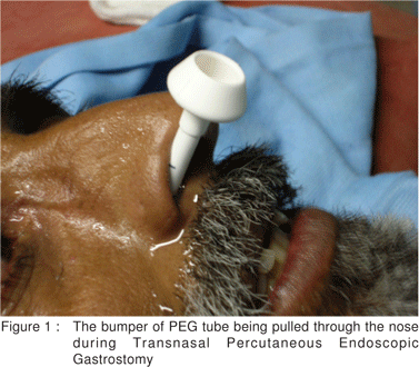
The nasal cavity was examined for deviated nasal septum or any other structural abnormality. Pre-operative preparation, like for conventional PEG, included shaving of upper anterior abdominal wall and administration of prophylactic antibiotic (Inj Amoxycillin – Calvulanic acid 1.2 gm IV). A cotton pledget soaked in 5% Lignocaine and 1 in 10000 Adrenaline was placed in one nostril 15 minutes prior to the procedure. Transnasal endoscope (Olympus GIF XQ 160 , length 110 cm, outer diameter 5.9 mm and channel diameter of 2 mm) was then passed and complete upper gastro-intestinal (GI) endoscopic examination was carried out. TPEG (PEG 18,Wilson Cook,18 F) was then performed by using the conventional pull technique under continuous cardiac and oxygen saturation monitoring. The bumper of the PEG device was pulled in and anchored under endoscopic guidance (Figure 1). The procedure lasted 10 minutes and 40 seconds from endoscopic intubation to anchoring of PEG device and was well tolerated. Feeding through the PEG was commenced after 24 hours and antibiotics were continued for 5 days. No long term complications were noted. The patient improved with chemo-radiotherapy and started accepting oral feeds after 10 weeks .The PEG device was removed 12 weeks after the procedure.
Discussion
The transnasal route for GI endoscopy was first described in 1987.[1] The role of diagnostic transnasal GI endoscopy was prospectively evaluated and minor adverse effects like selflimiting epistaxis (2.3%), nasal pain (1.6%) and vasovagal reaction (0.3%) were described.[2] This led to the use of transnasal GI endoscopy for placement of feeding tubes and PEG.[3] It has been estimated that in about 6% to 9% of patients with neurological disorders, trauma or tumours of the head and neck, it is not possible to carry out PEG by the oral route.[4] We have been using transnasal endoscopy for patients with corrosive ingestion, upper GI bleeding in a setting of cervical spine injuries and trismus and for endoscopic placement of nasogastric tube.

TPEG has minor modifications over the conventional oral procedure. The PEG device chosen should have a diameter of 16F to 18 F tube with a collapsible bumper. The bumper when compressed with the fingers should not be larger than the diameter of the endoscope. Although it is said that sedation may not be used for this procedure, a bolus of midazolam is preferable. The use of adrenaline with topical anaesthesia decreases the incidence of epistaxis. There is a theoretical risk of increased peristomal infection with organisms colonosing the nostrils. Use of Mupirocin ointment prior to the procedure has been advocated to decrease the colonization of Staphylococci. Prior evaluation of nostrils by an otorhinolaryngologist to select one with a larger passage diameter and absence of any obstructing lesion is also desirable. Following the bumper with the endoscope to ensure its proper expansion and placement has also been recommended. [5] This procedure allows PEG in patients with dysphagia where the oral route is not accessible. It also allows unsedated placement of PEG tube in high risk groups where sedation is not desirable. In fact, advocates of TPEG consider it to be the procedure of choice over conventional PEG due to increased patient tolerance and decreased side effects.[6] Although standard diameter gastroscopes have been used for TPEG, failure rates are likely to be higher, especially in patients with compromised nasal cavity.[7]
References
1. Johnson DA, Cattau EL Jr, Khan A, Newell DE, Chobanian SJ. Fibreoptic esophagogastroscopy via nasal intubation.Gastrointest Endosc. 1987;33:32–33.
2. Dumortier J, Ponchon T, Scoazec JY, Moulinier B. Zarka F, Paliard P, et al. Prospective evaluation of transnasal esophagogastroduodenoscopy: feasibility and study on performance and tolerance. Gastrointest Endosc. 1999;49:285– 91.
3. Kulling D, Bauerfeind P, Fried M. Transnasal versus transoral endoscopy for the placement of nasoenteral feeding tube in critically ill patients. Gastrointest Endosc. 2000;52:506–10.
4. Gibson SE, Wenig BL, Watkins JL. Complications of Percutaneous endoscopic gastrostomy in head and neck cancer patients. Ann Otol Rhinol Laryngol. 1992;101:46–50.
5. Vitale MA, Villotti G, D’Alba L, De Cesare MA, Frontespezi S, Iacopini G. Unsedated transnasal percutaneous endoscopic gastrostomy placement in selected patients. Endoscopy. 2005;37:48–51.
6. Dumortier J, Lapalus MG, Pereira A, Lagarrigue JP, Chavaillon A, Ponchon T. Unsedated transnasal PEG placement. Gastrointest Endosc. 2004;59:54–7.
7. Desai D, Joshi A, Abraham P, Chawla M, Sonsale A. Transnasal percutaneous endoscopic gastrostomy using a standarddiameter gastroscope (9.8mm). Endoscopy 2005;37:1035.